. 2010 Jun 17;115(24):5080-8.
doi: 10.1182/blood-2009-08-239020. Epub 2010 Mar 31.

The IgG-specific endoglycosidase EndoS inhibits both cellular and complement-mediated autoimmune hemolysis

Affiliations

The IgG-specific endoglycosidase EndoS inhibits both cellular and complement-mediated autoimmune hemolysis

Maria Allhorn et al. Blood. .

Abstract

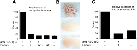
EndoS from Streptococcus pyogenes is an immunomodulating enzyme that specifically hydrolyzes glycans from human immunoglobulin G and thereby affects antibody effector functions. Autoimmune hemolytic anemia is caused by antibody-mediated red blood cell (RBC) destruction and often resists treatment with corticosteroids that also cause frequent adverse effects. We show here that anti-RhD (anti-D) and rabbit anti-human-RBC antibodies (anti-RBC) mediated destruction of RBC, ie, phagocytosis, complement activation, and hemolysis in vitro and in vivo was inhibited by EndoS. Phagocytosis by monocytes in vitro was inhibited by pretreatment of anti-D with EndoS before sensitization of RBCs and abrogated by direct addition of EndoS to blood containing sensitized RBCs. The toxic effects of monocytes stimulated with anti-D-sensitized RBCs, as measured by interleukin-8 secretion and oxygen metabolite production, was restrained by EndoS. Agglutination of RBCs and complement-mediated hemolysis in vitro in whole human blood caused by rabbit anti-RBCs was inhibited by EndoS. Development of anemia in mice caused by a murine anti-RBC immunoglobulin G2a monoclonal autoantibody and complement activation and erythrophagocytosis by Kupffer cells in the liver were reduced by EndoS. Our data indicate that EndoS is a potential therapeutic agent that might be evaluated as an alternative to current treatment regimens against antibody-mediated destruction of RBCs.


Figures

Figure 1
Figure 1
Treatment of anti-D with EndoS does not affect the binding of anti-D to RBCs. (A) Western blot analysis of anti-D on RBCs using peroxidase-conjugated anti–human IgG. (B) Glycan hydrolysis of anti-D on RBCs determined by lectin ELISA (optical density [OD] = 450 nm). (C) Quantification of RBC-bound anti-D by ELISA (OD = 450 nm) using antiserum against human IgG.
Figure 2
Figure 2
RBCs sensitized with anti-D IgG pretreated with EndoS are not attached to, or phagocytosed by, monocytes. (A) The degree of adherence/phagocytosis of RBCs sensitized with anti-D determined by monocyte monolayer assay and expressed as a number of monocytes with one or more adherent/phagocytosed RBCs of 100 monocytes detected. (B) The appearance of “rosettes” showed using Grünwald-Giemsa staining of monocyte monolayers incubated with RBCs sensitized with anti-D (top panel) and anti-D pretreated with EndoS (bottom panel; original magnification ×60). (C) Phagocytosis of sensitized RBCs by THP-1 cells and blood monocytes with or without treatment of anti-D with EndoS. The results are presented as the relative amount of internalized hemoglobin in cells detected by reactivity with 2,7-diaminofluorene. (D) Resuspended monocytes after incubation with RBCs sensitized with anti-D followed by the removal of noningested RBCs. (Left) RBCs sensitized with anti-D. (Right) Anti-D pretreated with EndoS. (E). Purified blood monocytes were incubated with FITC-labeled RBCs sensitized with anti-D and the degree of ingested RBCs determined by measuring the fluorescence.
Figure 3
Figure 3
EndoS inhibits metabolic oxygen responses induced by RBCs sensitized with anti-D immune plasma. Metabolic oxygen responses from monocytes were analyzed by measuring the chemiluminescence after addition of RBC-sensitized anti-D-positive plasma (with or without treatment with EndoS) and luminol, at 5-minute intervals for 1 hour. y-axis represents opsonic index, ie, the ratio of monocyte responses to sensitized RBCs versus unsensitized RBCs. control RBC indicates RBCs sensitized with nonimmune plasma.
Figure 4
Figure 4
EndoS inhibits secretion of IL-8 by monocytes induced by RBCs sensitized with anti-D. Relative quantification of IL-8 secretion in supernatants from THP-1 cells or blood monocytes incubated with RBCs sensitized with anti-D for 3 hours, with or without pretreatment of anti-D with EndoS. (Inset) IL-8 incubated with EndoS followed by separation on SDS-PAGE and staining with Coomassie Blue.
Figure 5
Figure 5
Hydrolysis of anti–human RBC-IgG by EndoS. (A) Rabbit antiserum against human RBCs was treated with EndoS or PBS and used for sensitization of RBCs. RBC-bound IgG was determined by solubilization of sensitized RBCs followed by Western blot using antiserum rabbit IgG. (B) Glycan hydrolysis of RBC-bound IgG determined by lectin ELISA (OD = 450 nm). (C) RBC-bound antibody was quantified by ELISA (OD = 450 nm) using antiserum against rabbit IgG.
Figure 6
Figure 6
EndoS inhibits hemolysis and agglutination of sensitized RBCs and prevents binding of C1q to sensitized RBCs. (A) Relative amount of hemoglobin quantified by 2,7-diaminofluorene in plasma after incubation of human blood with antiserum against human RBCs, pretreated with EndoS or not. (1)Direct addition of EndoS to blood containing RBC antiserum. (2)EndoS alone added to blood. NS indicates not significant. (B) Blood smears. (Top panel) A total of 80 μg of anti-RBC IgG added to 700 μL human blood, diluted 35 times in PBS, and incubated 1 hour at 37°C. (Middle panel) Anti-RBC treated with EndoS before addition to blood. (Bottom panel) No addition of antibodies. (C) Relative C1q deposition onto IgG-sensitized RBCs detected using FITC-conjugated anti–human C1q.
Figure 7
Figure 7
EndoS decreases the development of AIHA in BALB/c mice pretreated with 34-3C anti-RBC mAb. BALB/c mice were intravenously injected with 34-3C anti-RBC mAb; and 24 hours later, 20 μg of EndoS was intravenously injected. (A) Hematocrit values of individual mice treated with EndoS (○) or PBS (●) were measured 1, 2, 4, 6, and 8 days after injection of 200 μg 34-3C IgG2b mAb. Horizontal lines indicate mean values. (B). Representative histologic appearance of iron deposits in Kupffer cells from mice injected with 34-3C IgG2b anti-RBC mAb and then with EndoS. Note substantial reduction of iron deposits in livers from mice treated with EndoS, compared with completely untreated mice (original magnification ×200). (Top right panel) Higher magnification of iron deposits as indicated by arrows. (C) BALB/c mice were intravenously injected with 300 μg of 34-3C IgG1 or 200 μg of 34-3C IgG2a class-switch variant; and 24 hours later, 20 μg of EndoS was intravenously injected. Hematocrit values of individual mice treated with EndoS (○) or PBS (●) were measured 4 days after injection of 34-3C mAb. Horizontal lines indicate mean values. The normal range of hematocrit values (mean ± 3 SD) of 2- to 3-month-old BALB/c mice is represented as a shaded area.

References

    1. Petz L, Garatty G. Immune Hemolytic Anemias. 2nd ed. Philadelphia, PA: Churchill Livingstone; 2004.
    1. Kavai M, Szegedi G. Immune complex clearance by monocytes and macrophages in systemic lupus erythematosus. Autoimmun Rev. 2007;6(7):497–502. - PubMed
    1. Daniels G. Naming blood groups and the genes that control them. ISBT Science Series. 2009;4(1):118–120.
    1. Suto Y, Ishikawa Y, Hyodo H, Uchikawa M, Juji T. Gene organization and rearrangements at the human Rhesus blood group locus revealed by fiber-FISH analysis. Hum Genet. 2000;106(2):164–171. - PubMed
    1. Kumpel BM, Goodrick MJ, Pamphilon DH, et al. Human Rh D monoclonal antibodies (BRAD-3 and BRAD-5) cause accelerated clearance of Rh D+ red blood cells and suppression of Rh D immunization in Rh D− volunteers. Blood. 1995;86(5):1701–1709. - PubMed

Publication types

MeSH terms