Comparative Study
. 2010 May 1;184(9):5213-23.
doi: 10.4049/jimmunol.0902879. Epub 2010 Mar 31.

B cells have distinct roles in host protection against different nematode parasites

Affiliations
Comparative Study

B cells have distinct roles in host protection against different nematode parasites

Qian Liu et al. J Immunol. .

Abstract

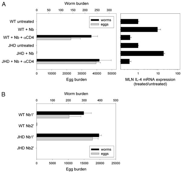
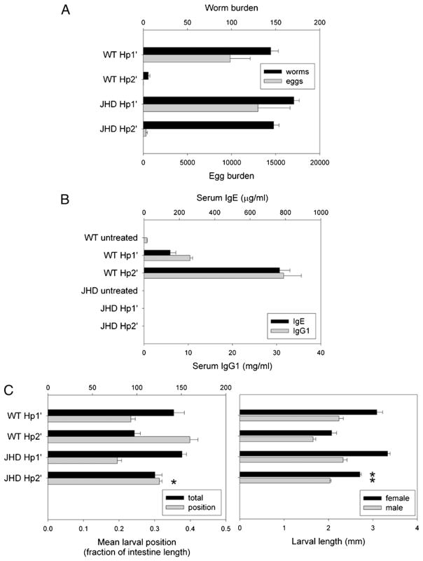
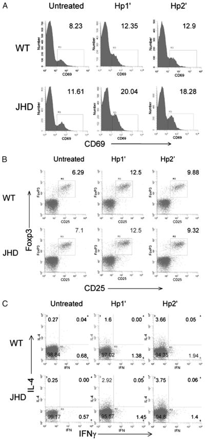
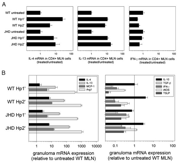
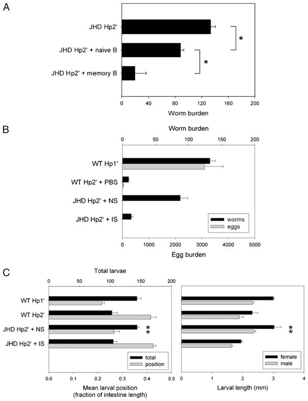
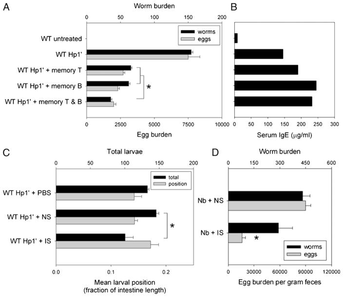
B cells can mediate protective responses against nematode parasites by supporting Th2 cell development and/or by producing Abs. To examine this, B cell-deficient mice were inoculated with Nippostrongylus brasiliensis or Heligmosomoides polygyrus. B cell-deficient and wild type mice showed similar elevations in Th2 cytokines and worm expulsion after N. brasiliensis inoculation. Worm expulsion was inhibited in H. polygyrus-inoculated B cell-deficient mice, although Th2 cytokine elevations in mucosal tissues were unaffected. Impaired larval migration and development was compromised as early as day 4 after H. polygyrus challenge, and administration of immune serum restored protective immunity in B cell-deficient mice, indicating a primary role for Ab. Immune serum even mediated protective effects when administered to naive mice prior to inoculation. This study suggests variability in the importance of B cells in mediating protection against intestinal nematode parasites, and it indicates an important role for Ab in resistance to tissue-dwelling parasites.


Conflict of interest statement

Disclosures

The authors have no financial conflicts of interest.

Figures

FIGURE 1
FIGURE 1
The development of Th2 cells in draining MLNs after N. brasiliensis inoculation does not require B cells. JHD and WT mice were inoculated s.c. with 500 N. brasiliensis L3 and sacrificed 10 d (A) or 7 d later (B–C). A, Frequency of B220+ cells in MLNs was analyzed by flow cytometry. Serum IgE levels were determined by ELISA. Results are representative of two independent experiments. B, The number of IL-4–producing cells in 1 million MLN cells was determined by ELISPOT assay. IL-4, IL-13, and IFN-γ mRNA expression in MLN cells were detected by real-time quantitative PCR. C, CD4+ T cells were isolated from MLN cells of N. brasiliensis-inoculated and untreated JHD and WT mice, and quantitative PCR was used to measure IL-4 and IL-13 mRNA. Results are representative of two independent experiments.
FIGURE 2
FIGURE 2
The host protective response to N. brasiliensis inoculation depends on CD4 cells but not B cells. A, JHD and WT mice were inoculated s.c. with N. brasiliensis L3, and some mice were administered an additional 1 mg anti-CD4 mAb once per week starting the day before inoculation. Mice were sacrificed 12 d postinoculation, and worm burden in small intestine and egg burden in large intestine were measured. IL-4 mRNA expression of MLN was detected by real-time quantitative PCR. B, JHD and WT mice were primed with N. brasiliensis L3 s.c. Two months later, these mice were given a secondary challenge (Nb2′) and compared with naive mice given a primary inoculation (Nb1′). At day 7 after challenge, mice were sacrificed and assayed for worm burden in the small intestine and egg burden in the large intestine. Results are representative of two independent experiments.
FIGURE 3
FIGURE 3
The host protection to acute or chronic H. polygyrus infection is B cell-dependent. JHD and WT mice were orally inoculated with 200 H. polygyrus L3, 2 wk later treated with antihelminthic, and then four weeks after drug cure rechallenged with H. polygyrus (Hp2′). Other mice were given primary challenge alone (Hp1′). A and B, The mice were sacrificed at day 14 after final inoculation. Worms and eggs were counted in the small intestinal lumen and fecal contents, respectively (A), and serum IgE and IgG1 levels were determined by ELISA (B). Results are representative of three independent experiments. C, Primary and secondary challenged mice were sacrificed at day 4 postinoculation. Tissue-dwelling larvae were counted in situ to determine total number and distribution along small intestine. Individual larvae were removed, sexed, and measured. Means represent at least 10 larvae per mouse, 4 mice per group. Asterisks mark significant difference (p <0.01) from WT Hp2′ group.
FIGURE 4
FIGURE 4
T cell activation, Treg cell frequency, and Th2 differentiation in draining MLNs were comparable in H. polygyrus-inoculated JHD and WT mice. Primary (Hp1′) and secondary (Hp2′) H. polygyrus challenges were administered to WT and JHD mice as described in Fig. 3. On day 7 after challenge, the mice were sacrificed, MLNs were collected, and cell suspensions were prepared. A, The cells were stained for anti-CD4 and anti-CD69. The expression of CD69 on CD4+ populations is shown. B, Cell suspensions were stained for surface CD4 and CD25 expression and intracellular Foxp3. Cells were gated on CD4+ population, and the frequency of CD25+ Foxp3+ Treg cells is shown. C, Cells were stimulated with PMA and inomycin for 6 h and stained for surface CD4 and intracellular IL-4 and IFN-γ. The IL-4 and IFN-γ production by CD4+ T cells is shown.
FIGURE 5
FIGURE 5
The cytokine profile at the host–parasite interface is intact in JHD mice. WT and JHD mice were challenged with primary and secondary H. polygyrus inoculation as previously described. A, CD4 T cells were purified from MLNs by positive selection and RNA was isolated. IL-4, IL-13, and IFN-γ mRNA expression by these CD4 T cells were detected by quantitative PCR. Results are representative of two independent experiments. B, On day 7 after challenge, granulomas were collected and RNA was isolated. IL-4, IL-13, MCP-1, Arg1, IL-10, TGF-β, IFN-γ, iNOS, and thymic stromal lymphopoietin mRNA expression were detected by real-time quantitative PCR. Results are representative of two independent experiments.
FIGURE 6
FIGURE 6
B cells and serum can restore the protective memory response against acute or chronic H. polygyrus infection in JHD mice. IS, NS, and memory B cells were collected from H. polygyrus-challenged WT mice. Naive B cells were isolated from untreated WT mice. JHD mice were primed with H. polygyrus and drug-cured in preparation for secondary challenge. B cells were transferred i.v. 2 d before secondary challenge, or serum was transferred i.p. every 3 d beginning 1 d before secondary challenge. A, Parasite burden at day 14 postinoculation was compared between groups of JHD Hp2′ mice given memory B, naive B, or no cells. B, Day 14 worm and egg burdens were compared for JHD Hp2′ with IS or NS versus WT and primary control groups. C, Day 4 larval burden, distribution, and development was measured as previously described for JHD Hp2′ given IS or NS. Results are representative of two independent experiments. Asterisks mark significant difference (p <0.01) from WT Hp2′ group, or as indicated by brackets.
FIGURE 7
FIGURE 7
T cells, B cells, and serum from immunized mice can transfer protection against acute or chronic primary H. polygyrus infection. WT mice were primed and drug-cured with H. polygyrus as described. Two months later, some mice were sacrificed for harvest of T and B cells (memory T, memory B). Other mice were rechallenged with H. polygyrus inoculation and exsanguinated at day 14 after secondary inoculation with IS. NS from untreated mice and PBS were used as control interventions. A and B, Memory B and/or T cells were transferred i.v. into naive WT mice 2 d before primary H. polygyrus inoculation. Worm and egg burden (A) and serum IgE (B) were assayed 14 d later as measurements of the response to chronic infection. C, WT mice were inoculated with H. polygyrus on day 0, administered PBS, NS, or IS on days – 1 and 2, and the distribution of tissue-dwelling larvae (L4) was observed as an indicator of the early immune response. D, WT mice were inoculated with N. brasiliensis on day 0, administered NS or IS on day – 1, and examined for worm burden on day 7 after inoculation. Asterisks mark significant difference (p <0.01) from the NS group, or as indicated by brackets.

Similar articles

Cited by

References

    1. Hotez PJ, Brindley PJ, Bethony JM, King CH, Pearce EJ, Jacobson J. Helminth infections: the great neglected tropical diseases. J Clin Invest. 2008;118:1311–1321. - PMC - PubMed
    1. Diemert DJ, Bethony JM, Hotez PJ. Hookworm vaccines. Clin Infect Dis. 2008;46:282–288. - PubMed
    1. Scales HE, Ierna MX, Lawrence CE. The role of IL-4, IL-13 and IL-4Ralpha in the development of protective and pathological responses to Trichinella spiralis. Parasite Immunol. 2007;29:81–91. - PubMed
    1. Finkelman FD, Shea-Donohue T, Morris SC, Gildea L, Strait R, Madden KB, Schopf L, Urban JF., Jr Interleukin-4- and interleukin-13-mediated host protection against intestinal nematode parasites. Immunol Rev. 2004;201:139–155. - PubMed
    1. Anthony RM, Rutitzky LI, Urban JF, Jr, Stadecker MJ, Gause WC. Protective immune mechanisms in helminth infection. Nat Rev Immunol. 2007;7:975–987. - PMC - PubMed

Publication types

MeSH terms