. 2010 Apr 8;464(7290):932-6.
doi: 10.1038/nature08944. Epub 2010 Mar 31.

The kinetics of two-dimensional TCR and pMHC interactions determine T-cell responsiveness

Affiliations

The kinetics of two-dimensional TCR and pMHC interactions determine T-cell responsiveness

Jun Huang et al. Nature. .

Abstract

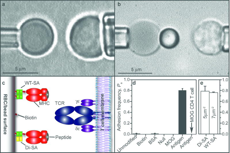
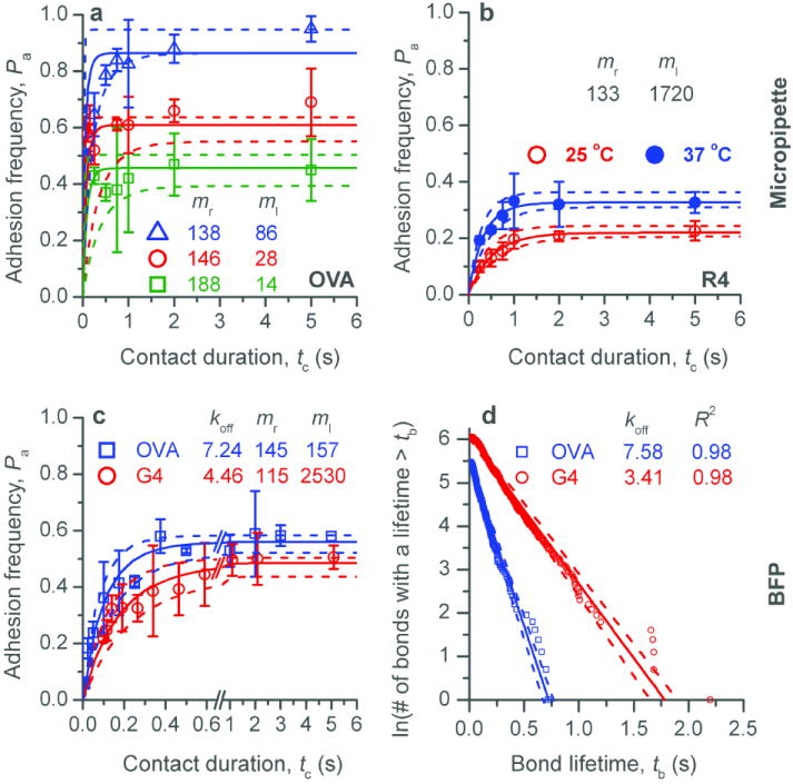
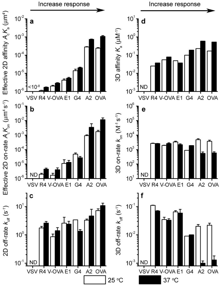
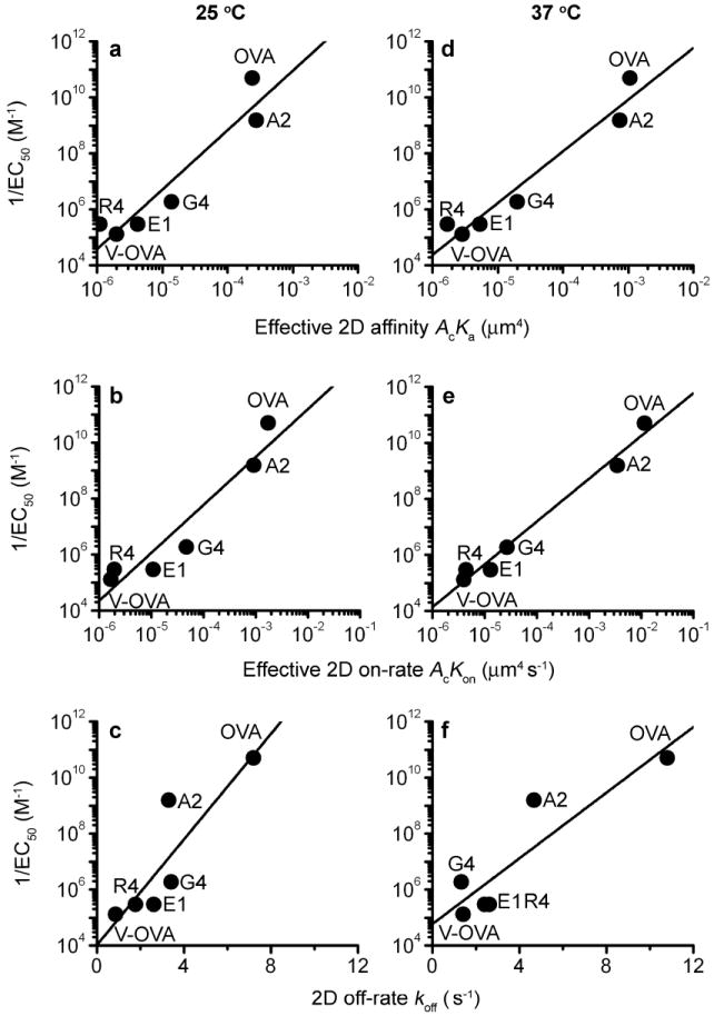
The T-cell receptor (TCR) interacts with peptide-major histocompatibility complexes (pMHC) to discriminate pathogens from self-antigens and trigger adaptive immune responses. Direct physical contact is required between the T cell and the antigen-presenting cell for cross-junctional binding where the TCR and pMHC are anchored on two-dimensional (2D) membranes of the apposing cells. Despite their 2D nature, TCR-pMHC binding kinetics have only been analysed three-dimensionally (3D) with a varying degree of correlation with the T-cell responsiveness. Here we use two mechanical assays to show high 2D affinities between a TCR and its antigenic pMHC driven by rapid on-rates. Compared to their 3D counterparts, 2D affinities and on-rates of the TCR for a panel of pMHC ligands possess far broader dynamic ranges that match that of their corresponding T-cell responses. The best 3D predictor of response is the off-rate, with agonist pMHC dissociating the slowest. In contrast, 2D off-rates are up to 8,300-fold faster, with the agonist pMHC dissociating the fastest. Our 2D data suggest rapid antigen sampling by T cells and serial engagement of a few agonist pMHCs by TCRs in a large self pMHC background. Thus, the cellular environment amplifies the intrinsic TCR-pMHC binding to generate broad affinities and rapid kinetics that determine T-cell responsiveness.


Figures

Figure 1
Figure 1
Micropipette and BFP. a and b, Micrographs of the micropipette (a) and BFP (b). A T cell (right) aspirated by a pipette was aligned with a RBC held stationary by another pipette (left) without (a, Movie M1) or with (b, Movies M2 and M3) a bead attached to the apex. c, RBCs or beads (left) were coupled by WT-SA or Di-SA with monomeric pMHC to interact with the TCR on T cells (right). d, Specificity controls of adhesion frequency measured at 5 s between OT1 T cells and unmodified RBCs, biotinylated RBCs without coating, biotinylated RBCs coated with BSA, null pMHC-I (VSV:H-2Kb), pMHC-II (MOG:I-Ab) or antigenic pMHC-I (OVA:H-2Kb), or between MOG CD4+ T cells and biotinylated RBCs coated with OVA:H-2Kb. e, Comparison between adhesion frequencies measured at 2 s using 7 and 5 μm-2 pMHC respectively captured by WT-SA and Di-SA.
Figure 2
Figure 2
2D kinetics measurements. a and b, Adhesion curves for the OT1 TCR interacting with OVA (a) and R4 (b) measured by micropipette at 25 °C (a) or both 25 and 37 °C (b) at indicated site densities. c, Adhesion curves of the OT1 TCR interacting with OVA and G4 measured by BFP at 25 °C. The data (points) were fitted (color matched solid curves) by a model for 2D binding kinetics (a, b and c). d, Pooled ensembles of 239 (OVA) or 424 (G4) lifetimes of bonds between the OT1 TCR and OVA (□) or G4 (○) were respectively sorted according to their durations. For each peptide, the natural log of the number of events with a lifetime ≥ tb was plotted vs. tb and fitted by a straight line. The negative slope represents off-rate koff (indicated). The goodness-of-fit was indicated by the R2 values. Color matched dotted curves represent 95% confidence intervals of the best-fit curves obtaining by bootstrapping.
Figure 3
Figure 3
Comparison of 2D and 3D kinetics. Affinities (a and d), on-rates (b and e), and off-rates (c and f) of the OT1 TCR interacting with indicated pMHCs. The 2D data (a-c) were measured by the adhesion frequency assay and analyzed with a monomeric binding model. The 3D data (d-f) from Refs. 11,12 were measured by surface plasmon resonance and analyzed with the same monomeric binding model except for the OVA and A2 data at 37 °C, which were analyzed with a dimeric binding model (values of the second-step kinetics were plotted).
Figure 4
Figure 4
Correlation between 2D kinetics and T cell proliferation. The reciprocal concentration required to reach half-maximal T cell proliferation (1/EC50) is plotted vs the effective 2D affinity (a and d), on-rate (b and e), and off-rate (c and f) measured at 25 °C (a-c) or 37 °C (d-f) for the indicated peptides. To quantify T cell proliferation, naïve OT1 splenocytes (3×10⁵/well) were cultured in 96-well plates with the indicated peptides at 37°C. After 48 h, 0.4 μCi/well of [3H] thymidine was added. After another 18 h, cells were harvested on a FilterMate harvester (PerkinElmer) and analyzed on a Matrix 96 Direct Beta Counter (PerkinElmer). EC50 values were calculated using GraphPad Prism.

Similar articles

Cited by

References

    1. Dustin ML, Bromley SK, Davis MM, Zhu C. Identification of self through two-dimensional chemistry and synapses. Annu Rev Cell Dev Biol. 2001;17:133–157. - PubMed
    1. Davis MM, et al. Ligand recognition by alpha beta T cell receptors. Annu Rev Immunol. 1998;16:523–544. - PubMed
    1. Gascoigne NR, Zal T, Alam SM. T-cell receptor binding kinetics in T-cell development and activation. Expert Rev Mol Med 2001. 2001:1–17. - PubMed
    1. Stone JD, Chervin AS, Kranz DM. T-cell receptor binding affinities and kinetics: impact on T-cell activity and specificity. Immunology. 2009;126(2):165–176. - PMC - PubMed
    1. Chesla SE, Selvaraj P, Zhu C. Measuring two-dimensional receptor-ligand binding kinetics by micropipette. Biophys J. 1998;75(3):1553–1572. - PMC - PubMed

Publication types

MeSH terms