. 2010 May;17(5):608-13.
doi: 10.1038/nsmb.1796. Epub 2010 Mar 31.

Structure of a clade C HIV-1 gp120 bound to CD4 and CD4-induced antibody reveals anti-CD4 polyreactivity

Affiliations

Structure of a clade C HIV-1 gp120 bound to CD4 and CD4-induced antibody reveals anti-CD4 polyreactivity

Ron Diskin et al. Nat Struct Mol Biol. 2010 May.

Abstract

Strategies to combat HIV-1 require structural knowledge of envelope proteins from viruses in HIV-1 clade C, the most rapidly spreading subtype in the world. We present a crystal structure containing a clade C gp120 envelope. The structure, a complex between gp120, the host receptor CD4 and the CD4-induced antibody 21c, reveals that the 21c epitope involves contacts with gp120, a nonself antigen, and with CD4, an autoantigen. Binding studies using wild-type and mutant CD4 show that 21c Fab binds CD4 in the absence of gp120, and that binding of 21c to clade C and HIV-2 gp120s requires the crystallographically observed 21c-CD4 interaction. Additional binding data suggest a role for the gp120 V1V2 loop in creating a high-affinity, but slow-forming, epitope for 21c after CD4 binds. These results contribute to a molecular understanding of CD4-induced antibodies and provide the first visualization to our knowledge of a potentially autoreactive antibody Fab complexed with both self and nonself antigens.


Figures

Fig. 1
Fig. 1
Structure of CAP210–sCD4–21c and comparison with other gp120–sCD4–CD4i-Fab complexes. (a) Ribbon representations of the 21c Fab (heavy chain in pink; light chain in yellow) and sCD4 (green) complexed with the clade C CAP210 gp120 (gray surface). (b) Ribbon representation of the CAP210 core gp120 structure with variable loops and other features highlighted. (c) Comparison of the CAP210–sCD4–21c structure with structures of sCD4–CD4i-Fab complexes including clade B gp120s (PDB codes 1G9M5, 2QAD3, 2B4C4 for HXBc2–sCD4–17b, YU2–sCD4–412d, and JR-FL–sCD4–X5, respectively). Structures are shown in surface representation with the Fab heavy chain in pink, the light chain in yellow, sCD4 in green and gp120 in gray.
Fig. 2
Fig. 2
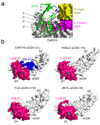
sCD4 interactions. (a) Close-up of the interaction between sCD4 D1 (green ribbons with letters referring to individual β-strands, blue highlights for 21c-contacting regions, and pink highlights for CAP210-contacting regions), CAP210 gp120 (gray), and the 21c heavy and light chains (pink and yellow, respectively). The sCD4 C’ strand is near the interface with gp120, but forms no contacts within 1.4 Å, creating a gap at the sCD4–gp120 interface as seen in previous structures,,. (b) Surface representations of sCD4 from different gp120–sCD4–CD4i-Fab structures (PDB codes: HXBc2–sCD4–17b: 1G9M5; YU2–sCD4–412d: 2QAD3; JR-FL–sCD4–X5: 2B4C4). sCD4 surface area that is buried at the interface with gp120 is highlighted in pink on each sCD4 structure with the number of Å2 buried on sCD4 by gp120 indicated. The CAP210–sCD4–21c structure is the only complex structure in which the CD4i Fab also contacts sCD4 (blue highlighted area along with the number of Å2 buried on sCD4 by 21c).
Fig. 3
Fig. 3
Comparison of CAP210 gp120 with clade B gp120 structures. The core CAP210 structure was superimposed with the HXBc2, YU2, and JR-FL structures (PDB codes 1G9M5, 2QAD3, 2B4C4 for HXBc2–sCD4–17b, YU2–sCD4–412d, and JR-FL–sCD4–X5, respectively) (top). The middle and bottom panels show superpositions of the V5 and CD4-binding loops. In the CAP210 V5 loop, the conserved N-linked site at Asn465 (comparable to Asn463 of YU2 to which carbohydrate is attached3,5) may not be glycosylated as the asparagine sidechain points to the interior of the V5 loop. The longer length of the V5 loop in CAP210 and other clade C gp120s as compared with clade B gp120s (Supplementary Fig. 1) may restrict access to the CD4 binding site.
Fig. 4
Fig. 4
Contact surfaces on 21c, sCD4, and gp120. (a) Surface representation of the 21c combining site (from the CAP210–sCD4–21c structure) with CDR1s in gold (H1) and cyan (L1); CDR2s in yellow (H2) and marine blue (L2); and CDR3s in red (H3) and dark blue (L3). (b) Footprints of gp120 (top) and sCD4 (bottom) on the 21c combining site. CDR contacts highlighted as in (a) with framework region contacts in pink. (c) 21c footprint on sCD4 (dark blue) and CAP210 (cyan).
Fig. 5
Fig. 5
21c interactions with sCD4 and gp120s. (a) Surface representation of CAP210–sCD4 (left) and CAP210–sCD4–21c (right) with a modeled carbohydrate at the location of the introduced N-linked glycosylation site (blue) in sCD4K75T. The introduced glycan is predicted to disrupt interactions with 21c, but is distant from the interface with gp120. (b) SDS-PAGE comparison of the mobility of wild-type sCD4 and sCD4K75T. The mobility shift to a higher apparent molecular weight of sCD4K75T compared with sCD4 is consistent with glycosylation at the introduced N-linked glycosylation site. (c) Equilibrium binding data (equilibrium binding response (Req) versus the log of the indicated protein concentrations) for SPR experiments in which sCD4, the sCD4K75T mutant, or CAP210 gp120 were injected over immobilized 21c. The best-fit binding curve to the experimental data is shown for the sCD4–21c interaction. (d) Sensorgrams from SPR experiments in which a concentration series (from 1.0 µM–31.2 nM, two-fold dilutions) of the indicated core gp120 protein (clade C CAP210 or HIV-2 UC1) was injected together with 2 µM sCD4 (orange curves) or sCD4K75T (blue curves, thickened for clarity) over immobilized 21c Fab (reproduced with residual plots in Supplementary Fig. 5). (e) Sensorgrams from SPR experiments in which a concentration series (from 1.0 µM–31.2 nM, two-fold dilutions) of the indicated gp120 protein containing the V1V2 loop (CAP210V1V2 or UC1Full-length) was injected together with 2 µM sCD4 (orange curves) or sCD4K75T (blue curves) over immobilized 21c Fab (reproduced with residual plots in Supplementary Fig. 5).

References

    1. UNAIDS. Report on the global HIV/AIDS epidemic. 2008
    1. McCutchan FE. Understanding the genetic diversity of HIV-1. AIDS. 2000;14(Suppl 3):S31–S44. - PubMed
    1. Huang CC, et al. Structures of the CCR5 N terminus and of a tyrosine-sulfated antibody with HIV-1 gp120 and CD4. Science. 2007;317:1930–1934. - PMC - PubMed
    1. Huang CC, et al. Structure of a V3-containing HIV-1 gp120 core. Science. 2005;310:1025–1028. - PMC - PubMed
    1. Kwong PD, et al. Structures of HIV-1 gp120 envelope glycoproteins from laboratory-adapted and primary isolates. Structure. 2000;8:1329–1339. - PubMed

Publication types