Smoking-Induced Acute Eosinophilic Pneumonia in a 15-year-old Girl: A Case Report
- PMID: 20358030
- PMCID: PMC2846739
- DOI: 10.4168/aair.2010.2.2.144
Smoking-Induced Acute Eosinophilic Pneumonia in a 15-year-old Girl: A Case Report
Abstract
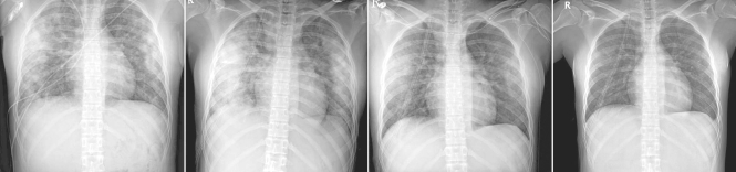
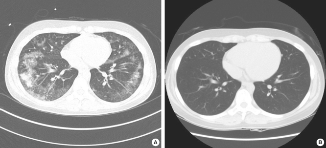
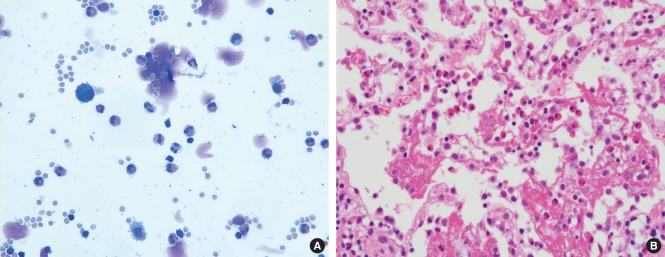
Acute eosinophilic pneumonia is a very rare disease that is characterized by acute febrile respiratory failure, diffuse bilateral infiltrates on chest X-ray, and eosinophilia in bronchoalveolar lavage fluid in the absence of infection. We present the case of a 15-year-old girl diagnosed with smoking-induced acute eosinophilic pneumonia. A previously healthy young girl with a 1-day history of fever presented with cough, dyspnea, and diffuse bilateral infiltrates on chest X-ray. She had started smoking only 3 weeks before presentation. She was diagnosed by bronchoalveolar lavage fluid tests and lung biopsy and dramatically improved after steroid treatment. We emphasize that acute eosinophilic pneumonia must be considered when acute pneumonia does not respond to broad-spectrum antibiotics. Effective treatment and prompt institution of therapy can obviate unnecessary morbidity and mortality.
Keywords: Pulmonary eosinophilia; adolescent; bronchoalveolar lavage fluid; smoking.
Conflict of interest statement
There are no financial or other issues that might lead to conflict of interest.
Figures
References
-
- Badesch DB, King TE, Jr, Schwarz MI. Acute eosinophilic pneumonia: a hypersensitivity phenomenon? Am Rev Respir Dis. 1989;139:249–252. - PubMed
-
- Allen JN, Pacht ER, Gadek JE, Davis WB. Acute eosinophilic pneumonia as a reversible cause of noninfectious respiratory failure. N Engl J Med. 1989;321:569–574. - PubMed
-
- Oermann CM, Panesar KS, Langston C, Larsen GL, Menendez AA, Schofield DE, Cosio C, Fan LL. Pulmonary infiltrates with eosinophilia syndromes in children. J Pediatr. 2000;136:351–358. - PubMed
-
- Pope-Harman AL, Davis WB, Allen ED, Christoforidis AJ, Allen JN. Acute eosinophilic pneumonia. A summary of 15 cases and review of the literature. Medicine (Baltimore) 1996;75:334–342. - PubMed
-
- Cottin V, Cordier JF. Eosinophilic pneumonias. Allergy. 2005;60:841–857. - PubMed