. 2010 Jul;67(14):2491-506.
doi: 10.1007/s00018-010-0351-5. Epub 2010 Apr 1.

Endocytosis of hepatitis C virus non-enveloped capsid-like particles induces MAPK-ERK1/2 signaling events

Affiliations

Endocytosis of hepatitis C virus non-enveloped capsid-like particles induces MAPK-ERK1/2 signaling events

Konstantina Katsarou et al. Cell Mol Life Sci. 2010 Jul.

Abstract

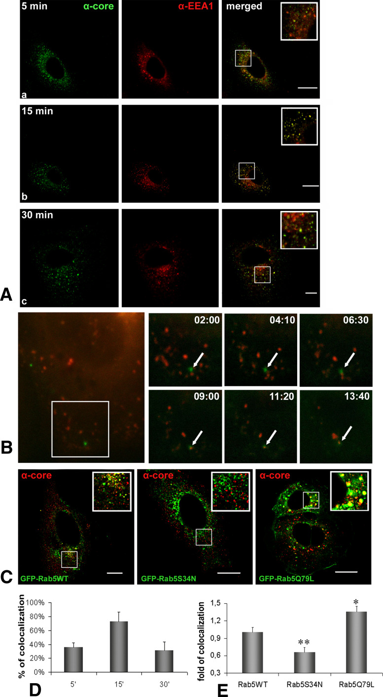
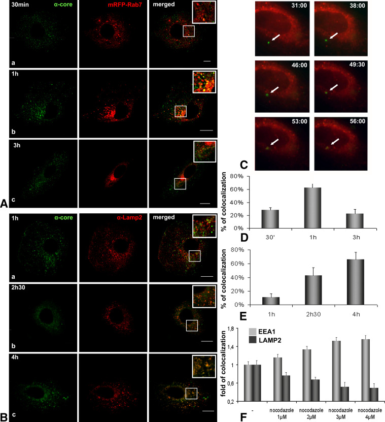
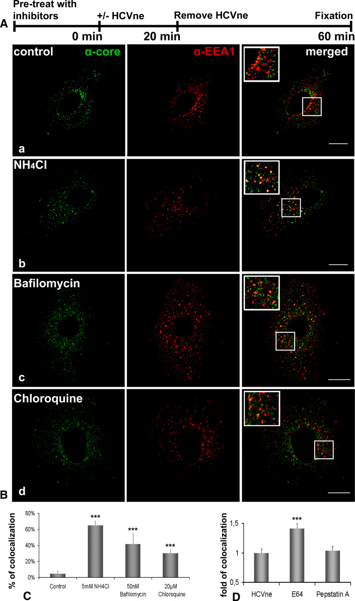
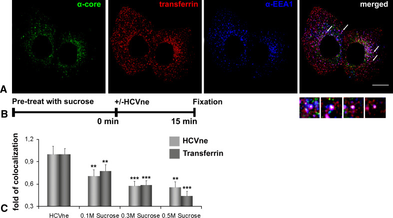
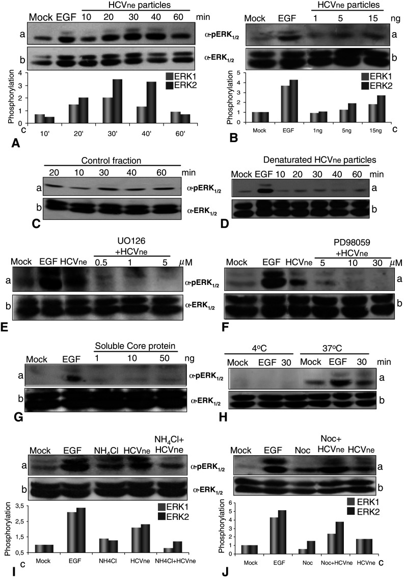
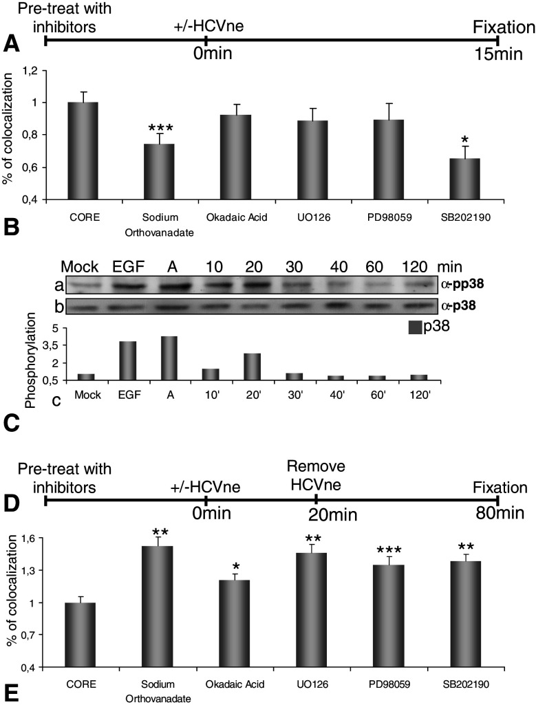
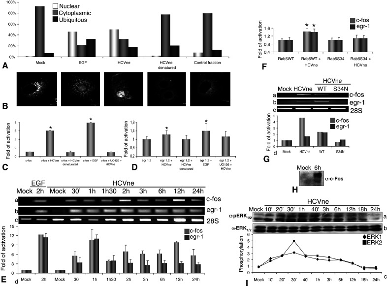
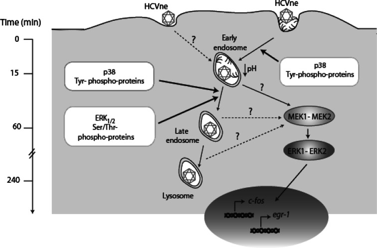
Although HCV is an enveloped virus, naked nucleocapsids have been reported in the serum of infected patients. The HCV core particle serves as a protective capsid shell for the viral genome and recombinant in vitro assembled HCV core particles induce strong specific immunity. We investigated the post-binding mechanism of recombinant core particle uptake and its intracellular fate. In hepatic cells, these particles are internalized, most likely in a clathrin-dependent pathway, reaching early to late endosomes and finally lysosomes. The endocytic acidic milieu is implicated in trafficking process. Using specific phosphoantibodies, signaling pathway inhibitors and chemical agents, ERK(1/2) was found to be activated in a sustained way after endocytosis, followed by downstream immediate early genes (c-fos and egr-1) modulation. We propose that the intriguing properties of cellular internalization of HCV non-enveloped particles can induce specific ERK(1/2)-MAPKs events that could be important in HCV life cycle and pathogenesis of HCV infection.


Figures

Fig. 1
Fig. 1
HCVne particles enter early endosomes. A Confocal microscopy of immunolabeled Huh7 cells incubated with HCVne particles using anti-core (green) or anti-EEA1 (red) antibodies at 5 (a), 15 (b), or 30 (c) min. Colocalization is shown in yellow in the merged images and inset. Βars a , b 20 μm, c 8 μm. B Selected images obtained from time lapse videos (Movie S1), representing time trajectories of movement of GFP fluorescent HCVne particles [14] to early endosomes in mRFP-Rab5 transfected cells. Arrows indicate traffic of fluorescent spot (min:s). C Huh7 transfected cells with GFP-Rab5WT, GFP-Rab5S34N, or GFP-Rab5Q79L plasmids treated with HCVne particles for 15 min. Immunofluorescence staining with anti-core (red) antibody. Bars a 20 μm, b,c 16 μm. D,E Image-Pro Plus quantification of colocalization for HCVne with EEA1 from (A) and with GFP-Rab5 WT and mutants from (C) (the colocalization level measured in GFP-Rab5 WT was used as the basis of comparison). *P < 0.05, **P < 0.01
Fig. 2
Fig. 2
Presence of HCVne particles in late endosomes and lysosomes. A Confocal microscopy using anti-core (green) antibody and mRFP-Rab7 plasmid (red) for 30 min (a), 1 (b), and 3 (c) h of HCVne particles incubation. Colocalization is observed in yellow in the merged images and inset. Bars a 8 μm, b 16 μm, c 20 μm. B Following HCVne particles addition, Huh7 cells, were co-labelled with anti-core (green) and anti-Lamp2 (red) antibodies at 1 (a), 2.5 (b), or 4 (c) h of incubation at 37°C. Insets show high magnification regions of the merged images. Bars 16 μm. C Selected images obtained from time lapse videos (ESM, Movie 2), representing time trajectories of movement of GFP fluorescent HCVne particles [14] to late endosomes in mRFP-Rab7 expressing cells. Arrows indicate traffic of fluorescent HCVne particles (min:s). D,E Colocalization’s quantification for HCVne particles with Rab7 and Lamp-2, respectively. F Disruption of microtubules with increasing concentrations of nocodazole. Cells were pretreated for 2 h, HCVne were added for 20 min, and cells were fixed 4 h later. EEA1-HCVne colocalization is shown in light grey bars and Lamp2-HCVne in dark grey bars
Fig. 3
Fig. 3
HCVne particles require low pH for internalization. A Schematic diagram representation of the experiment. B Huh7 cells untreated (a) or incubated with 5 mM NH4Cl (b), 20 μM chloroquine (c), or 50 nM bafilomycin (d) were added with particles as described in (A) and immunolabeled with anti-core (green) and anti-EEA1 (red) antibodies. Inset images show a higher magnification of the boxed section of the merged pictures. Bars a,b,d 16 μm, c 20 μm. C Image-Pro quantification of colocalization. The control was arbitrarily set at 100% and all other values are a percentage of this. D Quantification of colocalization from cells treated with E64 (50 μm, 2.5 h) and pepstatin A (50 μm, 2.5 h). Cells were pretreated with the inhibitors as described in (A). ***P < 0.001
Fig. 4
Fig. 4
HCVne particles are internalised via a clathrin-dependent pathway. A HCVne particles and Alexa 546-transferrin (red) in Huh7 cells, processed for immunofluorescence with anti-core (green) and anti-EEA1 (blue) antibodies. Arrows show individual endosomes positive for the two markers (insets). Bar 16 μm. B Schematic diagram representation of the experiment. C Quantification of colocalization of HCVne particles and transferrin with EEA1. Huh7 cells pre-treated with increasing concentrations of sucrose. Following addition of HCVne particles or transferrin, cells were fixed and immunostained either with anti-core/anti-EEA1 or anti-EEA1. **P < 0.01, ***P < 0.001
Fig. 5
Fig. 5
HCVne particles activate MEK1/2–ERK1/2 pathway. A Time course of ERK1/2 phosphorylation from lysates of HepG2 cells maintained in serum-free conditions incubated with HCVne particles (5 ng) for 10, 20, 30, 40, and 60 min at 37°C. EGF (50 nM) was used as positive control. Western blot with phospho-ERK1/2 (a), ERK1/2 (b), and quantification of the optical densities of phospho-ERK immunoreactive bands normalized to the optical densities of total ERK1/2 in the same samples (c) are presented. ERK1 is represented in light grey bars and ERK2 in dark grey bars. B HepG2 cells, maintained in serum-free conditions, were incubated for 30 min with increasing concentrations of HCVne particles (expressed in ng of core protein). C HepG2 cells treated with a fraction of equivalent sucrose density from Sf9 cell lysates infected with a control (GFP only producing) baculovirus or D with the HCVne fraction heat denatured (1 h at 100°C). E,F HepG2 cells were pre-treated with MEK inhibitors UO126 or PD98059 at increasing concentrations before being treated with HCVne particles for 30 min. G HepG2 cells treated with soluble bacterial core protein added at increasing concentrations for 30 min. H HepG2 cells were incubated at either 4°C or 37°C followed by 30 min of HCVne particles or 10 min of EGF addition. I,J NH4Cl (5 mM, 15 min) or nocodazole (4 μM, 2 h) treated cells with HCVne for 30 min. All lysates were analyzed by immunoblotting as described in A
Fig. 6
Fig. 6
Implication of host cell proteins in HCVne trafficking. A,D Schematic representation of the experiment. HepG2 cells were pre-treated with sodium orthovanadate (5 mM, 30 min), okadaic acid (0.5 μM, 30 min), UO126 (1 μM, 1.5 h), PD98059 (5 mM, 1.5 h), and SB202190 (20 μM, 1.5 h). Particles were added for either 15 min (B) or for 20 min, removed and incubated until 80 min (E). Cells were fixed, immunostained with anti-core (green) and anti-EEA1 (red) and observed with confocal microscopy. Then, 9–15 confocal sections from three to five different experiments were selected and quantified with Image-Pro Plus software. C HCVne particles were added in HepG2 cells for indicated times. EGF (50 nM) and anisomycin labeled (A, 10 μM) were used as positive controls. Lysates were analysed by western blotting with anti-phospho-p38 (a) or anti-p38 (b) antibodies. Densitometric analysis is expressed in arbitrary units (c). *P < 0.05, **P < 0.01, ***P < 0.001
Fig. 7
Fig. 7
Endocytosis-dependent ERK1/2 nuclear translocation, sustained activation and IEG upregulation. A Untreated starved Huh7 cells or treated with EGF (50nM, 10 min), HCVne particles (30 min), heat denaturated fraction (1 h at 100°C, 30 min), and a fraction of equivalent sucrose density from Sf9 cell lysates infected with a control (GFP producing) baculovirus (30 min) were fixed and immunostained with anti-ERK1/2. Then, 100 cells were counted for nuclear and cytoplasmic ERK localisation. B Representative confocal sections are presented. CF HepG2 cells were transfected with pFos WT-GL3 (c-fos), Egr1.2-luc (egr-1), and/or GFP-Rab5WT, GFP-Rab5S34N plasmids for 24 h, then serum starved for 8 h, and treated or not with HCVne particles or with controls (same fraction heat denatured) for 18 h. Relative light units were measured and values were normalized to the total protein amount. E,G Total mRNAs from HepG2 cells transfected or not with GFP-Rab5WT, GFPRab5S34N were isolated at indicated time points and cells incubated with HCVne. RT–PCR was performed with specific primers for c-fos (a), egr-1 (b), 28S (c). Densitometric results were normalized against 28S are in arbitrary units (c) (representative experiment of triplicates). H Nuclear extract of HepG2 cells treated for 6 h with HCVne particles and immunostained with c-fos antibody. I Incubation of cells with HCVne particles at various times, immunostained with anti-phospho ERK1/2 (a), ERK1/2 (b), and densitometric analysis of phospho-ERK1 and ERK2 after normalisation against total ERKs is also presented in (c). *P < 0.05
Fig. 8
Fig. 8
Hypothetical model for HCVne particle entry HCVne particles enter the cells most likely by the clathrin-dependent pathway, even though an alternative entry pathway is not excluded, and reach early endosomes. p38 as well as tyrosine phosphatases are involved in this process. At 1 h of incubation, particles are progressing to late endosomes in a ERK1/2 and serine/threonine phosphatases-dependent manner. Finally, particles reach the lysosomes (4 h). During this progression, the MEK1/2–ERK1/2 pathway is activated in a sustainable manner. When phosphorylated, ERKs proteins translocate to the nucleus and activate immediate early genes c-fos and egr-1

Similar articles

Cited by

References

    1. But DY, Lai CL, Yuen MF. Natural history of hepatitis-related hepatocellular carcinoma. World J Gastroenterol. 2008;14:1652–1656. doi: 10.3748/wjg.14.1652. - DOI - PMC - PubMed
    1. Andre P, Perlemuter G, Budkowska A, Brechot C, Lotteau V. Hepatitis C virus particles and lipoprotein metabolism. Semin Liver Dis. 2005;25:93–104. doi: 10.1055/s-2005-864785. - DOI - PubMed
    1. Aoyagi K, Ohue C, Iida K, Kimura T, Tanaka E, Kiyosawa K, Yagi S. Development of a simple and highly sensitive enzyme immunoassay for hepatitis C virus core antigen. J Clin Microbiol. 1999;37:1802–1808. - PMC - PubMed
    1. Maillard P, Krawczynski K, Nitkiewicz J, Bronnert C, Sidorkiewicz M, Gounon P, Dubuisson J, Faure G, Crainic R, Budkowska A. Nonenveloped nucleocapsids of hepatitis C virus in the serum of infected patients. J Virol. 2001;75:8240–8250. doi: 10.1128/JVI.75.17.8240-8250.2001. - DOI - PMC - PubMed
    1. Bouvier-Alias M, Patel K, Dahari H, Beaucourt S, Larderie P, Blatt L, Hezode C, Picchio G, Dhumeaux D, Neumann AU, McHutchison JG, Pawlotsky JM. Clinical utility of total HCV core antigen quantification: a new indirect marker of HCV replication. Hepatology. 2002;36:211–218. doi: 10.1053/jhep.2002.34130. - DOI - PubMed

Publication types

MeSH terms

Substances

LinkOut - more resources