Review
. 2010 Aug;67(16):2717-47.
doi: 10.1007/s00018-010-0346-2. Epub 2010 Apr 1.

Retroviral reverse transcriptases

Affiliations
Review

Retroviral reverse transcriptases

Alon Herschhorn et al. Cell Mol Life Sci. 2010 Aug.

Abstract

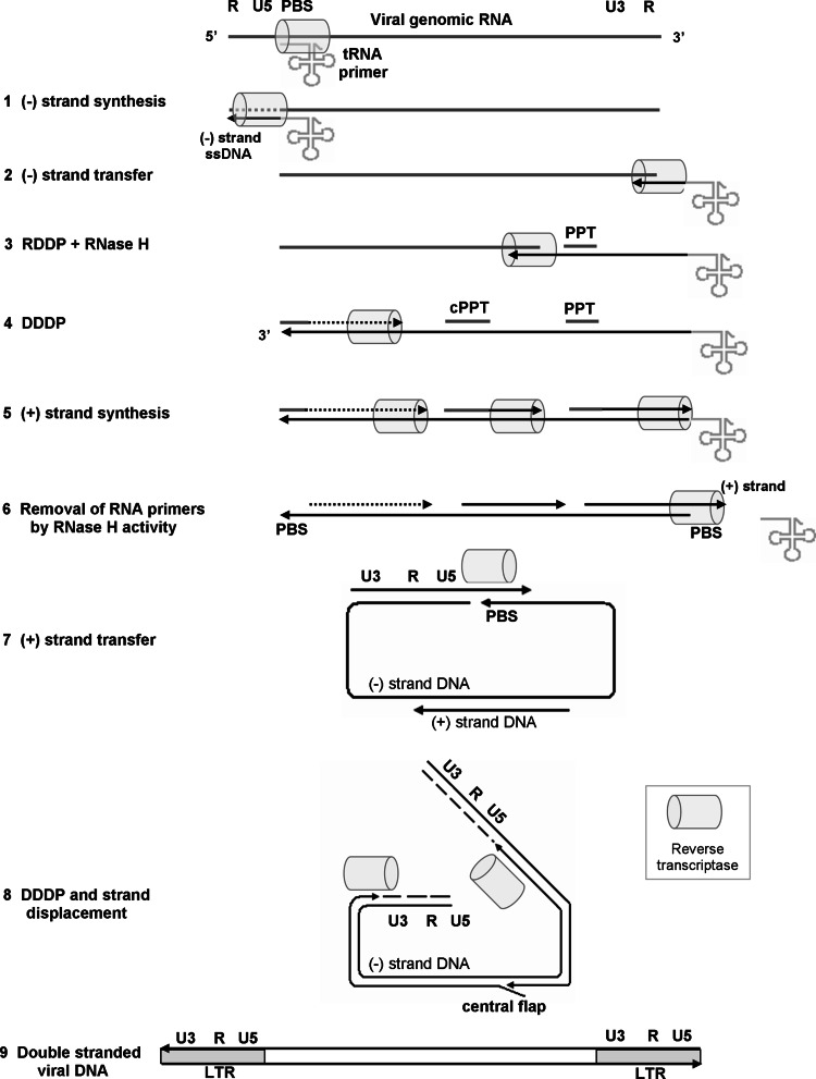
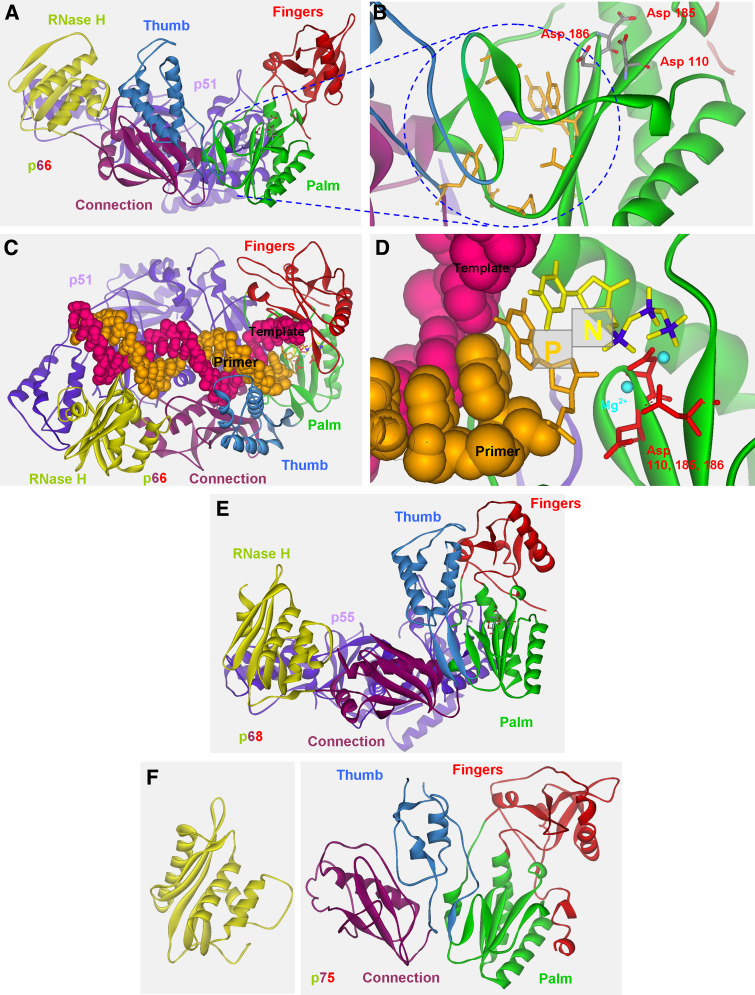
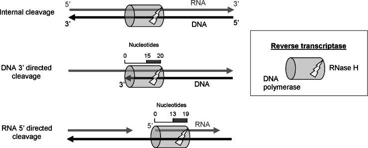
Reverse transcription is a critical step in the life cycle of all retroviruses and related retrotransposons. This complex process is performed exclusively by the retroviral reverse transcriptase (RT) enzyme that converts the viral single-stranded RNA into integration-competent double-stranded DNA. Although all RTs have similar catalytic activities, they significantly differ in several aspects of their catalytic properties, their structures and subunit composition. The RT of human immunodeficiency virus type-1 (HIV-1), the virus causing acquired immunodeficiency syndrome (AIDS), is a prime target for the development of antiretroviral drug therapy of HIV-1/AIDS carriers. Therefore, despite the fundamental contributions of other RTs to the understanding of RTs and retrovirology, most recent RT studies are related to HIV-1 RT. In this review we summarize the basic properties of different RTs. These include, among other topics, their structures, enzymatic activities, interactions with both viral and host proteins, RT inhibition and resistance to antiretroviral drugs.


Figures

Fig. 1
Fig. 1
The biogenesis of RTs in selected retroviral groups. Retroviral RTs are cleaved from the Gag-Pro-Pol polyprotein precursor. In lentiviruses the Gag-Pro-Pol polyprotein is synthesized after a single translational frameshift event. a In HIV-1, the resulting RT, which is cleaved from the polyprotein, is a heterodimer with the molecular weight of p66/p51. b In MMTV, the Gag-Pro-Pol is generated by two subsequent events of translational frameshift. The resulting RT is a monomer with 603 amino acids, of which 576 derived from the pol gene and 27 from the C-terminal portion of the Pro protein. Therefore, there is a putative internal cleavage site (IC) at this position within the Pro. The Gag-Pro polyprotein is generated by a single frameshift event and may be also the source for all the Gag and Pro derived proteins. c MLV uses a completely different strategy in which the Gag-Pro-Pol polyprotein is generated by an in-frame suppression of the stop codon. In the case of MLV, the scheme shows the protein lengths of the Moloney strain. This schematic description was not drawn to scale
Fig. 2
Fig. 2
The reverse transcription process. A schematic description of the different steps of the RTN process, drawn not to scale. See the text for a detailed description. RNA is depicted in grey and DNA in black. The direction of DNA synthesis is marked by the arrowheads. In various retroviruses (+) DNA synthesis can potentially be initiated from multiple RNA primers. The initiation of (+) DNA synthesis from the cPPT and 3′-PPT is shown in this scheme as straight black arrows (step 7), as these are the main initiation sites of the RTN of HIV-1. An additional potential site is shown upstream as a dotted arrow
Fig. 3
Fig. 3
The 3-dimensional structures of HIV-1, HIV-2 and MLV RTs. a Spatial structure of wild-type HIV-1 RT (PDB entry 1FK9) with its defined subdomains. HIV-1 RT was co-crystallized with the inhibitor efavirenz and is displayed from the side view after removing the inhibitor. b Magnification of the NNRTI hydrophobic pocket from a along with the three aspartic acids that form the catalytic site of the DNA polymerase domain. a, b Adapted with permission from Herschhorn et al. [201]. c The catalytic complex of HIV-1 RT together with a primer-template and dTTP (PDB entry 1RTD) from a top view. RT is displayed as solid ribbon, the template-primer as CPK (Corey, Pauling, and Kultin), and the 3′ nucleotide of the primer as well as the dTTP are displayed as a stick model. d Magnification of the N and P sites (marked as N and P) adjacent to the DNA polymerase sites from c and their position in relation to the three Asp catalytic residues. In c and d a hydroxyl was added to the 3′ position of the ribose in the P site to illustrate the presence of dNTP during DNA synthesis (the original complex was co-crystallized with ddNTP nucleotide). e The structure of HIV-2 RT (PDB entry 1MU2). f The structure of MLV RT was resolved in detail as two fragments, one for DNA polymerase domain (PDB entry 1RW3, right side) and the second for the RNase H domain (PDB entry 2HB5, left side). In all panels, the fingers subdomain is depicted in red, palm subdomain in green, thumb subdomain in light blue, connection subdomain in purple and RNase H domain in yellow (all of the large subunit or the only subunit in the case of MLV). All structures were displayed with Accelrys Discovery Studio Visualizer v1.6 (Accelrys Software Inc.)
Fig. 4
Fig. 4
A schematic description of the three modes of RT-associated RNase H cleavage. In all heteroduplex substrates for RNase H activity, the RNA strand is depicted in gray and DNA strand in black, and in both strands the 3′ termini are indicated by arrowheads. The viral RNA or host tRNA can be cleaved in either internal sites (top), or relative to the recessed end of either the DNA or RNA (middle and bottom schemes, respectively). When the DNA polymerase domain is associated with the 3′-end of the DNA strand (middle), the cleavage window on the RNA is between 15 and 20 nts opposite to the 3′-end of the DNA strand. Alternatively, in the recessed RNA 5′-end cleavage mode the polymerase active site is positioned on the DNA strand opposite to or near the RNA 5′-end. In this case the cleavage window is between 13 and 19 nts
Fig. 5
Fig. 5
HIV-1 RT inhibitors and drug-resistant RT mutants. a Incorporation of AZT-TP into the nascent DNA strand by HIV-1 RT can lead to two alternatives. Translocation of the incorporated NRTI into the P site (P) may be followed by binding of the next dNTP to the N site, resulting in the formation of a dead-end complex and inhibition of any further DNA synthesis. Conversely, excision of the chain terminator, AZT-MP, which is mediated by ATP as a pyrophosphate donor, releases the incorporated NRTI and consequently reverses inhibition. b The three-dimensional structure of the incorporated AZT-MP at the end of the primer at either the N site (N, yellow) or the P site (P, cyan). The pre- and post-translocation structures of HIV-1 RT are from PDB entries 1N6Q and 1N5Y, respectively. The coordinates of AZT-MP at the P site were taken from the post-translocation structure and were used to insert this molecule into the pre-translocation structure. This was done to indicate the general orientation of AZT-MP, and it does not necessary represent the exact position of AZT-MP in the P site. Residues that are associated with increased resistance to AZT are displayed in CPK style and are labeled. The primer and template are displayed in line style and the AZT-MP in stick style (yellow for the N site and cyan for the P site). HIV-1 RT is displayed as a solid ribbon in green. c, d The hydrophobic pocket of HIV-1 RT that binds NNRTIs in two structures of RT mutants. HIV-1 RTs with different mutations, which are associated with resistance to classical NNRTIs, were co-crystallized with the TMC278 NNRTI, a broad-range inhibitor of many mutant RT variants. The double mutant L100I + K103N is shown in (c) and the double mutant K103N + Y181C in (d). The highly potent inhibitor is flexible and can accommodate and interact with different residue in different mutants. Inhibiting the K103N + Y181C double mutant is mediated at least partially by interacting with Y183 that leads to its movement and the repositioning by 1.5 Å in the conserved Y183 MDD [194]. Residues of the hydrophobic pocket are displayed in orange and the mutant ones in yellow. All residues are presented as stick model and labeled. The distance between Y183 and the cyano group of TMC278 in both structures is also shown. All structures were displayed with Accelrys Discovery Studio Visualizer v1.6 (Accelrys Software Inc.)

References

    1. Coffin JM, Hughes SH, Varmus HE. Retroviruses. Cold Spring Harbor: Cold Spring Harbor Laboratory Press; 1997. - PubMed
    1. Skalka AM, Goff SP. Reverse transcriptase. Cold Spring Harbor: Cold Spring Harbor Laboratory Press; 1993. - PubMed
    1. Menendez-Arias L, Berkhout B (2008) Special issue on: retroviral reverse transcription. Virus Res 134 - PubMed
    1. Parniak MA (2004) Special issue: molecular biology of HIV. Int J Biochem Cell Biol 36
    1. Katz RA, Skalka AM. The retroviral enzymes. Annu Rev Biochem. 1994;63:133–173. doi: 10.1146/annurev.bi.63.070194.001025. - DOI - PubMed

MeSH terms

LinkOut - more resources