Review
. 2010:100:1-29.
doi: 10.1007/978-3-7643-8338-1_1.

Toxins from bacteria

Affiliations
Review

Toxins from bacteria

James S Henkel et al. EXS. 2010.

Abstract

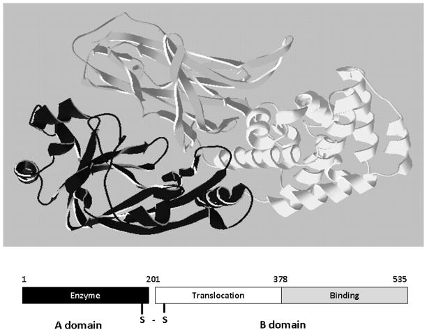
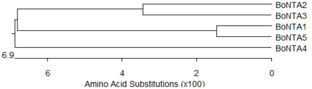
Bacterial toxins damage the host at the site of bacterial infection or distant from the site. Bacterial toxins can be single proteins or oligomeric protein complexes that are organized with distinct AB structure-function properties. The A domain encodes a catalytic activity. ADP ribosylation of host proteins is the earliest post-translational modification determined to be performed by bacterial toxins; other modifications include glucosylation and proteolysis. Bacterial toxins also catalyze the non-covalent modification of host protein function or can modify host cell properties through direct protein-protein interactions. The B domain includes two functional domains: a receptor-binding domain, which defines the tropism of a toxin for a cell and a translocation domain that delivers the A domain across a lipid bilayer, either on the plasma membrane or the endosome. Bacterial toxins are often characterized based upon the secretion mechanism that delivers the toxin out of the bacterium, termed types I-VII. This review summarizes the major families of bacterial toxins and also describes the specific structure-function properties of the botulinum neurotoxins.


Figures

Figure 1
Figure 1. AB organization of bacterial toxins
Diphtheria toxin is an AB toxin where the N terminal A domain (black) encodes an Enzyme activity, an ADP-ribosyl trasferase activity, NAD + EF-2 → ADP-r-EF-2 + nicotinamide + H+. The C-terminal B domain encodes a receptor binding function (grey) that binds to a grwoth factor receptor and enters cells through receptor-mediated endocytosis and a translocation function (white) which undergoes a pH-dependent conformation a change where chaafed amino acids are protonated which allows a pair of hydrophobic alpha helices to insert into the endosome membrane which is responsible for the delivery of A domain in to the host cytosol. Structure: PDB 1fol
Figure 2
Figure 2. Binding of superantigen SEC3 to TCR β
Staphylococcus aureus superantigen SEC3 binds to the β chain of TCR through the hypervariable domain 4 (HV4), acting as a wedge to encourage TCR and MHCII interaction and activation lacking foreign peptide specificity. Arrow indicates HV4 loop on TCR β. Structure: PDB 1jck.
Figure 3
Figure 3. Functional orgainzation of the type III cytotoxin Pseudomaons aeruginosa
ExoS is a bi-functional toxins and is orgainzed inot discret functional doamisn (amino acids): secretion domain (1–15), chaperone binding domain (16–51), membrane localization domain (51–77), Rho GAP domain, active site residue R146 (96–243), and ADP-ribosyltransferase domain, active site residues E379, E381 (233–453).
Figure 4
Figure 4. Schematic and structure of BoNT/A
A. Schematic of BoNT/A as an A/B toxin, linked by a disulfide bond between the light (LC) and heavy (HC) chains. B. Crystal structure of BoNT/A. LC (black) is the catalytically active domain and contains a HEXXH motif for coordination of the Zn2+ atom. The HC contains the N-terminal translocation (grey) domain and the C-terminal receptor-binding domain (light grey). Grey sphere represents zinc atom within LC active site. Colors between schematic and structure are coordinated with each domain. Structure: PDB 3bta.
Figure 5
Figure 5. Phylogenetic dendogram of BoNT/A1–A5 amino acid identity
Dendogram constructed using DNAStar® MegAlign.

Similar articles

Cited by

References

    1. Field M, GLH, Laird WJ, Smith PL. Heat-stable enterotoxin of Escherichia coli: In vitro effects on guanylate cyclase activity, cyclic GMP concentration, and ion transport in small intestine. Proc Natl Acad Sci. 1978;75:2800–2804. - PMC - PubMed
    1. Giannella RA. Pathogenesis of acute bacterial diarrheal disorders. Ann Rev Med. 1981;32:341–357. - PubMed
    1. Crane JK, WMS, Bolen EJ, Sando JJ, Linden J, Guerrant RL, Sears CL. Regulation of intestinal guanylate cyclase by the heat-stable enterotoxin of Escherichia coli (STa) and protein kinase C. Infect Immun. 1992;60:5004–5012. - PMC - PubMed
    1. Takao T, HT, Aimoto S, Shimonishi Y, Hara S, Takeda T, Takeda Y, Miwatani T. Amino acid sequence of a heat-stable enterotoxin isolated from enterotoxigentic Escherichia coli strain 18D. FEBS Lett. 1983;152:1–5. - PubMed
    1. Ozaki H, ST, Kubota H, Hata Y, Katsube Y, Shimonishi Y. Molecular structure of the toxic domain of heat-stable enterotoxin produced by enterotoxigenic Escherichia coli. J Biol Chem. 1991;266:5934–5941. - PubMed

Publication types

LinkOut - more resources