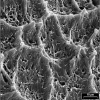
Variations in human DEJ scallop size with tooth type
- PMID: 20359518
- PMCID: PMC2880199
- DOI: 10.1016/j.jdent.2010.03.010
Variations in human DEJ scallop size with tooth type
Abstract
Objective: Recent literature suggests that the scalloped structure of the dentino-enamel junction (DEJ) is critical for DEJ stability. Aim of our study was to see if there are differences in scallop size and shape with tooth type.
Methods: Enamel of extracted permanent human teeth was demineralised using EDTA. After fixation and dehydration the scallops of the DEJ were investigated in a scanning electron microscope. Scallop area and shape (circularity) were measured for molars, premolars, canines and incisors.
Results: Scallop area showed main effects for tooth type and specimen, while, due to high variability in third molars, there was also an interaction effect (repeated measures two-way ANOVA, p<0.05). Differences between tooth types were statistically significant, suggesting that posterior teeth showed larger scallops compared to anterior teeth. Differences in shape (circularity) were not statistically significant.
Conclusion: Our results suggest that teeth which are subject to higher masticatory loads (posterior teeth) show larger and more pronounced scallops. These findings might be of interest for improving other interfaces joining dissimilar materials.
Copyright 2010 Elsevier Ltd. All rights reserved.
Figures


Similar articles
-
Insular dentin formation pattern in human odontogenesis in relation to the scalloped dentino-enamel junction.Ann Anat. 2007;189(3):243-50. doi: 10.1016/j.aanat.2006.11.007. Ann Anat. 2007. PMID: 17534031
-
Microstructure of primary tooth dentin.Pediatr Dent. 1999 Nov-Dec;21(7):439-44. Pediatr Dent. 1999. PMID: 10633518
-
Variable cementoenamel junction in one person.J Prosthet Dent. 1991 Jan;65(1):93-7. doi: 10.1016/0022-3913(91)90057-4. J Prosthet Dent. 1991. PMID: 2033555
-
Enamel pearls and cervical enamel projections on 2 maxillary molars with localized periodontal disease: case report and histologic study.Oral Surg Oral Med Oral Pathol Oral Radiol Endod. 2000 Apr;89(4):493-7. doi: 10.1016/s1079-2104(00)70131-4. Oral Surg Oral Med Oral Pathol Oral Radiol Endod. 2000. PMID: 10760733 Review.
-
Mesiodistal tooth size in non-syndromic unilateral cleft lip and palate patients: a meta-analysis.Clin Oral Investig. 2013 Mar;17(2):365-77. doi: 10.1007/s00784-012-0819-9. Epub 2012 Aug 23. Clin Oral Investig. 2013. PMID: 23011523 Review.
Cited by
-
Effect of interface surface design on the fracture behavior of bilayered composites.Eur J Oral Sci. 2019 Jun;127(3):276-284. doi: 10.1111/eos.12617. Epub 2019 Apr 19. Eur J Oral Sci. 2019. PMID: 31002749 Free PMC article.
-
Micromechanical interlocking structure at the filler/resin interface for dental composites: a review.Int J Oral Sci. 2023 May 31;15(1):21. doi: 10.1038/s41368-023-00226-3. Int J Oral Sci. 2023. PMID: 37258568 Free PMC article. Review.
References
-
- Imbeni V, Kruzic JJ, Marshall GW, Marshall SJ, Ritchie RO. The dentin-enamel junction and the fracture of human teeth. Nature Materials. 2005;4:229–32. - PubMed
-
- Dong XD, Ruse ND. Fatigue crack propagation path across the dentinoenamel junction complex in human teeth. Journal of Biomedical Materials Research Part A. 2003;66A:103–9. - PubMed
-
- Lin CP, Douglas WH, Erlandsen SL. Scanning electron-microscopy of type I collagen at the dentin-enamel junction of human teeth. Journal of Histochemistry & Cytochemistry. 1993;41:381–8. - PubMed
-
- Rasmussen ST. Fracture properties of human teeth in proximity to the dentinoenamel junction. Journal of Dental Research. 1984;63:1279–83. - PubMed
-
- Lin CP, Douglas WH. Structure-property relations and crack resistance at the bovine dentin-enamel junction. Journal of Dental Research. 1994;73:1072–8. - PubMed
Publication types
MeSH terms
Grants and funding
LinkOut - more resources
Full Text Sources

