

Next generation sequencing in research and diagnostics of ocular birth defects
- PMID: 20359920
- PMCID: PMC2871986
- DOI: 10.1016/j.ymgme.2010.03.004
Next generation sequencing in research and diagnostics of ocular birth defects
Abstract
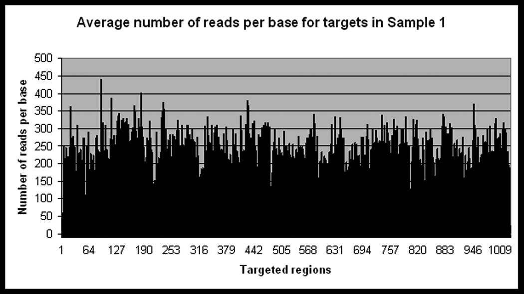
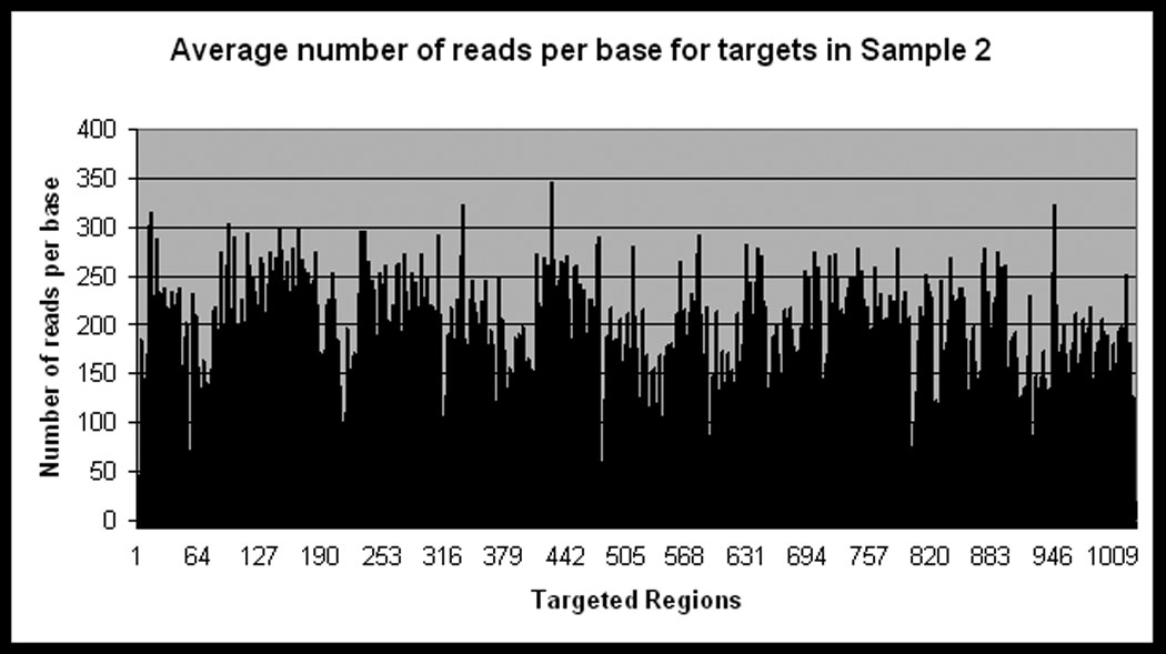
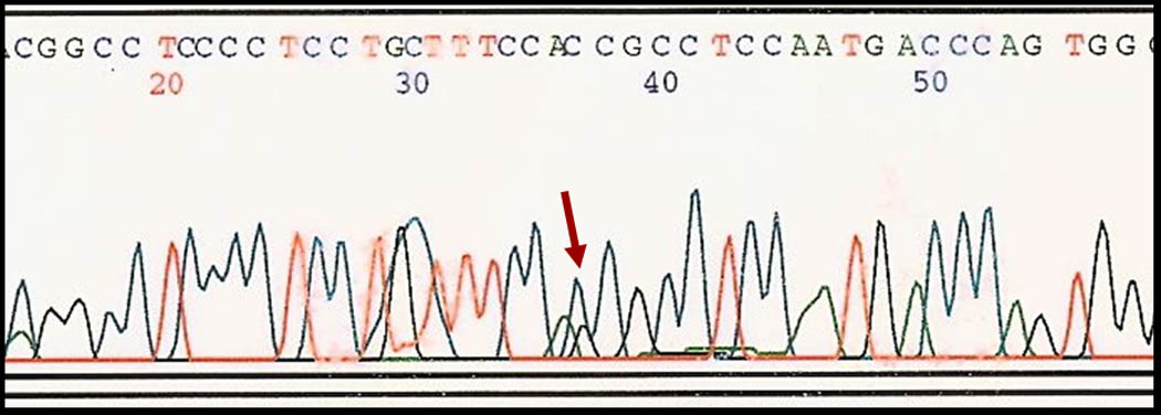
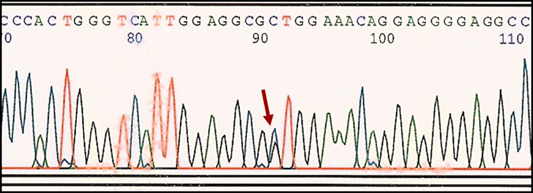
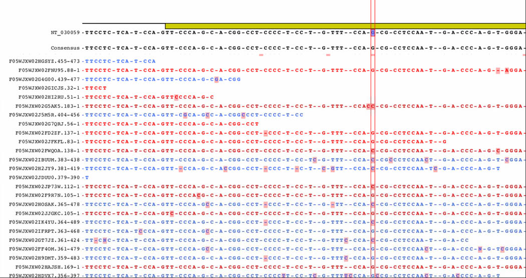
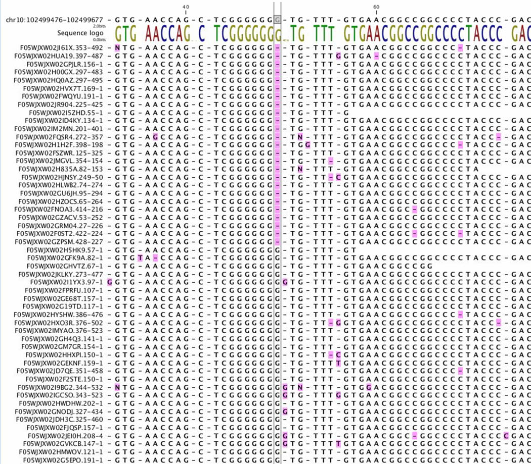
Sequence capture enrichment (SCE) strategies and massively parallel next generation sequencing (NGS) are expected to increase the rate of gene discovery for genetically heterogeneous hereditary diseases, but at present, there are very few examples of successful application of these technologic advances in translational research and clinical testing. Our study assessed whether array based target enrichment followed by re-sequencing on the Roche Genome Sequencer FLX (GS FLX) system could be used for novel mutation identification in more than 1000 exons representing 100 candidate genes for ocular birth defects, and as a control, whether these methods could detect two known mutations in the PAX2 gene. We assayed two samples with heterozygous sequence changes in PAX2 that were previously identified by conventional Sanger sequencing. These changes were a c.527G>C (S176T) substitution and a single basepair deletion c.77delG. The nucleotide substitution c.527G>C was easily identified by NGS. A deletion of one base in a long polyG stretch (c.77delG) was not registered initially by the GS Reference Mapper, but was detected in repeated analysis using two different software packages. Different approaches were evaluated for distinguishing false positives (sequencing errors) and benign polymorphisms from potentially pathogenic sequence changes that require further follow-up. Although improvements will be necessary in accuracy, speed, ease of data analysis and cost, our study confirms that NGS can be used in research and diagnostic settings to screen for mutations in hundreds of loci in genetically heterogeneous human diseases.
Figures
Similar articles
-
Evaluation of oligonucleotide sequence capture arrays and comparison of next-generation sequencing platforms for use in molecular diagnostics.Clin Chem. 2010 Aug;56(8):1297-306. doi: 10.1373/clinchem.2010.145441. Epub 2010 Jun 18. Clin Chem. 2010. PMID: 20562348
-
Assessment of target enrichment platforms using massively parallel sequencing for the mutation detection for congenital muscular dystrophy.J Mol Diagn. 2012 May-Jun;14(3):233-46. doi: 10.1016/j.jmoldx.2012.01.009. Epub 2012 Mar 16. J Mol Diagn. 2012. PMID: 22426012 Free PMC article.
-
The Genome Sequencer FLX System--longer reads, more applications, straight forward bioinformatics and more complete data sets.J Biotechnol. 2008 Aug 31;136(1-2):3-10. doi: 10.1016/j.jbiotec.2008.03.021. Epub 2008 Jun 21. J Biotechnol. 2008. PMID: 18616967 Review.
-
Targeted sequence capture and GS-FLX Titanium sequencing of 23 hypertrophic and dilated cardiomyopathy genes: implementation into diagnostics.J Med Genet. 2013 Sep;50(9):614-26. doi: 10.1136/jmedgenet-2012-101231. Epub 2013 Jun 19. J Med Genet. 2013. PMID: 23785128 Free PMC article.
-
An introduction to the informatics of "next-generation" sequencing.Curr Protoc Bioinformatics. 2011 Dec;Chapter 11:11.1.1-11.1.9. doi: 10.1002/0471250953.bi1101s36. Curr Protoc Bioinformatics. 2011. PMID: 22161566 Review.
Cited by
-
Detection of reassortant influenza B strains from 2004 to 2015 seasons in Barcelona (Catalonia, Spain) by whole genome sequencing.Virus Res. 2023 Jun;330:199089. doi: 10.1016/j.virusres.2023.199089. Epub 2023 Apr 5. Virus Res. 2023. PMID: 37011863 Free PMC article.
-
A novel application of pattern recognition for accurate SNP and indel discovery from high-throughput data: targeted resequencing of the glucocorticoid receptor co-chaperone FKBP5 in a Caucasian population.Mol Genet Metab. 2011 Dec;104(4):457-69. doi: 10.1016/j.ymgme.2011.08.019. Epub 2011 Aug 24. Mol Genet Metab. 2011. PMID: 21917492 Free PMC article.
-
High-throughput molecular diagnosis of von Willebrand disease by next generation sequencing methods.Haematologica. 2012 Jul;97(7):1003-7. doi: 10.3324/haematol.2011.055285. Epub 2012 Feb 7. Haematologica. 2012. PMID: 22315491 Free PMC article.
-
High-frequency, low-coverage "false positives" mutations may be true in GS Junior sequencing studies.Sci Rep. 2017 Oct 23;7(1):13751. doi: 10.1038/s41598-017-13116-6. Sci Rep. 2017. PMID: 29062110 Free PMC article.
-
SYK expression in monomorphic epitheliotropic intestinal T-cell lymphoma.Mod Pathol. 2018 Mar;31(3):505-516. doi: 10.1038/modpathol.2017.145. Epub 2017 Oct 20. Mod Pathol. 2018. PMID: 29052597
References
MeSH terms
Grants and funding
LinkOut - more resources
Full Text Sources
