. 2010 Jun 11;285(24):18769-80.
doi: 10.1074/jbc.M110.129007. Epub 2010 Apr 1.

Phospho-regulated ACAP4-Ezrin interaction is essential for histamine-stimulated parietal cell secretion

Affiliations

Phospho-regulated ACAP4-Ezrin interaction is essential for histamine-stimulated parietal cell secretion

Xia Ding et al. J Biol Chem. .

Abstract

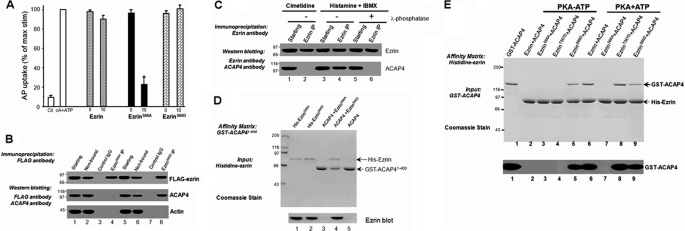
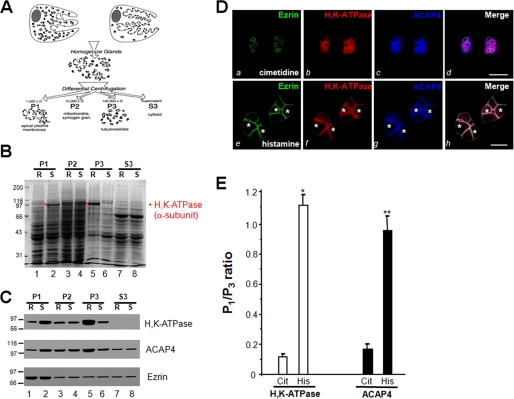
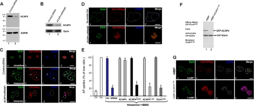
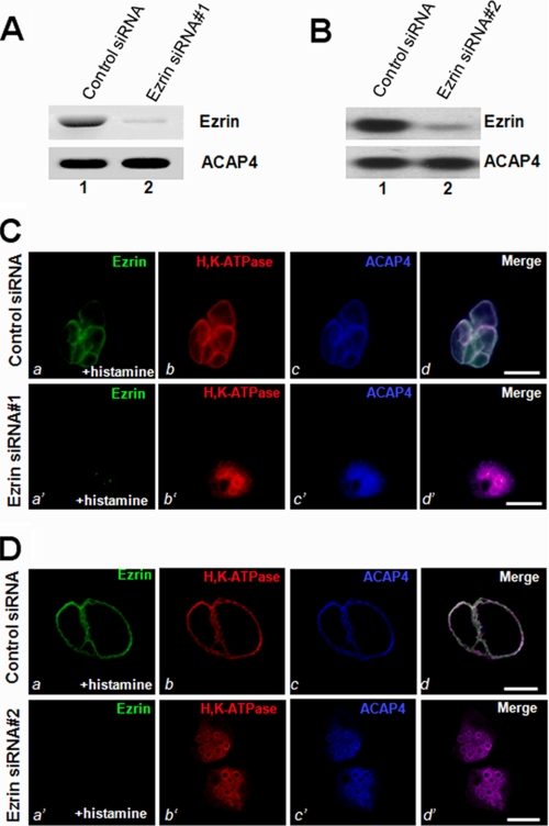
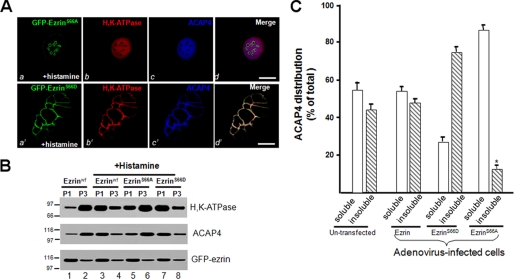
The ezrin-radixin-moesin proteins provide a regulated linkage between membrane proteins and the cortical cytoskeleton and also participate in signal transduction pathways. Ezrin is localized to the apical membrane of parietal cells and couples the protein kinase A activation cascade to the regulated HCl secretion. Our recent proteomic study revealed a protein complex of ezrin-ACAP4-ARF6 essential for volatile membrane remodeling (Fang, Z., Miao, Y., Ding, X., Deng, H., Liu, S., Wang, F., Zhou, R., Watson, C., Fu, C., Hu, Q., Lillard, J. W., Jr., Powell, M., Chen, Y., Forte, J. G., and Yao, X. (2006) Mol. Cell Proteomics 5, 1437-1449). However, knowledge of whether ACAP4 physically interacts with ezrin and how their interaction is integrated into membrane-cytoskeletal remodeling has remained elusive. Here we provide the first evidence that ezrin interacts with ACAP4 in a protein kinase A-mediated phosphorylation-dependent manner through the N-terminal 400 amino acids of ACAP4. ACAP4 locates in the cytoplasmic membrane in resting parietal cells but translocates to the apical plasma membrane upon histamine stimulation. ACAP4 was precipitated with ezrin from secreting but not resting parietal cell lysates, suggesting a phospho-regulated interaction. Indeed, this interaction is abolished by phosphatase treatment and validated by an in vitro reconstitution assay using phospho-mimicking ezrin(S66D). Importantly, ezrin specifies the apical distribution of ACAP4 in secreting parietal cells because either suppression of ezrin or overexpression of non-phosphorylatable ezrin prevents the apical localization of ACAP4. In addition, overexpressing GTPase-activating protein-deficient ACAP4 results in an inhibition of apical membrane-cytoskeletal remodeling and gastric acid secretion. Taken together, these results define a novel molecular mechanism linking ACAP4-ezrin interaction to polarized epithelial secretion.


Figures

FIGURE 1.
FIGURE 1.
Phosphorylation of Ser66 of ezrin specifies its association with ACAP4. A, non-phosphorylatable ezrinS66A inhibits acid secretion in SLO-permeabilized gastric glands. Glands were then stimulated with 100 μm cAMP plus 100 μm ATP, and the AP uptake was measured as described under “Materials and Methods.” AP data are plotted as a percentage of the stimulated control for each experiment. Error bars represent S.E.; n = 5. *, significant difference from stimulated controls (p < 0.05). B, co-precipitation of FLAG-ezrinS66D, but not FLAG-ezrinS66A, with ACAP4 from transfected parietal cell cultures, as described under “Materials and Methods.” Immunoprecipitates were fractionated by SDS-PAGE, followed by transferring onto nitrocellulose membrane for immunoblotting of FLAG-ezrin, ACAP, and actin. C, co-precipitation of endogenous ezrin-ACAP4 complex from stimulated, but not resting, gastric glands, as described under “Materials and Methods.” Dephosphorylation of ezrin with λ-phosphatase treatment disrupts ezrin-ACAP4 complex isolated from stimulated glands. D, ACAP4 binds to Ser66-phosphorylated ezrin via its N terminus. GST-ACAP4(1–400) was purified on glutathione-agarose beads and used as an affinity matrix for absorption of purified recombinant ezrin proteins (phospho-mimicking and non-phosphorylatable mutants). Top, SDS-polyacrylamide gel; bottom, Western blotting of ezrin. E, ACAP4-binding activity analysis of wild-type and mutant ezrin proteins in the presence of PKA alone (PKA-ATP; no phosphorylation) or PKA plus ATP (PKA + ATP; phosphorylation). Top, Coomassie Blue-stained SDS-polyacrylamide gel. ACAP4 binding was observed after incubation of ezrin with both PKA and ATP (lane 6) but not with PKA in the absence of ATP (lane 2). The addition of PKA on its own, without ATP, had no effect (not shown). In addition, the ACAP4 binding activity of ezrinS66A and ezrinS66D was unaffected by treatment of PKA and ATP (lanes 7 and 9). Bottom, Western blotting of ezrin.
FIGURE 2.
FIGURE 2.
Histamine stimulation triggers the redistribution of ACAP4 to the apical membrane of parietal cells. A, schematic diagram of subcellular fractionation of resting and stimulated gastric glands. B, SDS-PAGE of subcellular fractions from resting (R) and stimulated (S) gastric gland preparations. Equal total proteins (35 μg/lane) were used. C, Western blotting analyses of ezrin, ACAP4, and H,K-ATPase (α-subunit) of subcellular fractions derived from resting and stimulated gastric glands. Note that stimulation enriches the protein levels of H,K-ATPase and ACAP4 in P1 fraction. D, this montage represents confocal images collected from resting and secreting gastric parietal cells triply stained for ezrin (green), H,K-ATPase (red), ACAP4 (blue), and their merged images. Ezrin is mainly located in the apical plasma membrane of parietal cells (seen as rings) in a pattern suggestive of the apical plasma membrane invaginations that form the intracellular canaliculi (a). Labeling of ACAP4 is mainly located in the cytoplasm of parietal cells (c), which is mainly co-localized to that of H,K-ATPase distribution (b; red), whereas a lesser degree of co-localization is seen in apical membrane. Their apical localization becomes readily evident when the three images are merged (d; white). Bar, 15 μm. Stimulation induces remodeling of the apical membrane, seen as a dilation of apical vacuoles in the parietal cells (e–g). Labeling of ACAP4 is mainly located in the dilated apical vacuole membrane (g, asterisk), which is superimposed onto that of ezrin distribution in the merge (h). Bar, 15 μm. E, densitometric quantitation of the α-subunit of H,K-ATPase and ACAP4 proteins from P1 (plasma membrane-enriched) and P3 (tubulovesicle-enriched) fractions. The measurements were expressed as P1/P3 ratio. All data are given as means ± S.E. (error bars) of four preparations. Cit, cimetidine; His, histamine.
FIGURE 3.
FIGURE 3.
ACAP4 is essential for parietal cell acid secretion. A and B, parietal cells were transfected with the ACAP4 siRNA oligonucleotides for 48 h and subjected to SDS-PAGE and immunoblotting. Left, immunoblot for ACAP4; right, immunoblot for ezrin. Scrambled oligonucleotides were used as controls. C and D, these two sets of confocal images were collected from resting and secreting gastric parietal cells triply stained for ezrin (green), H,K-ATPase (red), and ACAP4 (blue). Ezrin is mainly located in the apical vacuole membrane of parietal cells, which was seen as rings in control siRNA-treated cells (a). Stimulation of control siRNA-treated parietal induces a dilation of apical vacuole membrane (a′). However, suppression of ACAP4 (siRNA 1 and siRNA 2) by siRNA abolishes the apical concentration of H,K-ATPase (f) but not ezrin (e). In addition, knockdown of ACAP4 attenuates the dilation of apical vacuoles (cversus g; circled). Bar, 15 μm. E, inhibition of parietal cell secretion by GAP-deficient ACAP4. Cultured parietal cells were infected by adenovirus-containing wild type and mutant ACAP4 for 2 h before stimulation with histamine and IBMX, and AP uptakes were measured. AP uptakes are shown for resting and stimulated controls and for stimulated glands treated with either full-length ACAP or ACAP4 mutants or ezrinS66D mutant. AP data are plotted as a percentage of the stimulated control for each experiment. Error bars, S.E. (n = 4 for all experiments). In a separate preparation, aliquots of cultured parietal cells were transfected with ACAP4 siRNA and scrambled oligonucleotides followed by a standardized AP uptake assay. *, p < 0.01 compared with stimulated controls. max stim, maximal stimulation. F, addition of ACAP4(1–400) recombinant protein disrupts ezrin-ACAP4 association. Purified GST-ezrinS66D protein on glutathione-Sepharose beads were used as an affinity matrix for absorbing GFP-ACAP4. After extensive washes, aliquots of ACAP4-bound GST-ezrinS66D-Sepharose beads were incubated with 5 μm purified ACAP4(1–400) protein on a rotator for 30 min at room temperature. After extensive washes, the Sepharose beads were boiled in SDS-PAGE sample buffer, and bound proteins were fractionated by SDS-PAGE, followed by visualization in Coomassie Blue stain. Note that incubation of ACAP4(1–400) peptide with ACAP4-bound GST-ezrinS66D-Sepharose beads disrupts ezrin-ACAP4 interaction. G, ezrin-ACAP4 is essential for apical translocation of ACAP4 and H,K-ATPase. To directly assess the role of ACAP4-ezrin interaction in parietal cell activation, recombinant ACAP4(1–400) protein was introduced into SLO-permeabilized cells, followed by the addition of cAMP/ATP. Twenty minutes after the treatment, cultured parietal cells were then fixed for immunocytochemistry. Stimulation of SLO-permeabilized cells with cAMP plus ATP induced a dilation of apical vacuole membrane in the presence of MBP protein (a–d). However, the addition of ACAP4(1–400) peptide abolishes the apical concentration of H,K-ATPase (f) and ACAP4 (g) but not ezrin (e). Bar, 15 μm.
FIGURE 4.
FIGURE 4.
Ezrin is essential for the apical localization of ACAP4 during parietal cell activation. A and B, RNA interference of ezrin. Cultured parietal cells were transfected with the ezrin siRNA oligonucleotides (siRNA 1 and 2) for 36 h and subjected to SDS-PAGE and immunoblotting. Top, immunoblot of ezrin; bottom, immunoblot for ACAP4. C and D, this set of optical images was collected from gastric parietal cells treated with ezrin RNA interference oligonucleotide (siRNA 1 and 2) and triply stained for ezrin (green), H,K-ATPase (red), and ACAP4 (blue). In ezrin siRNA-treated secreting cells, ACAP4 staining was diffused throughout the cytoplasm. The suppression of ezrin by siRNA abolishes the apical concentration of H,K-ATPase and subsequent failure of apical membrane vacuole dilation (b′; red). However, stimulation of control siRNA-treated parietal induces a dilation of apical vacuole membrane (a; green) and concentration of H,K-ATPase to the dilated apical vacuoles. Bar, 15 μm.
FIGURE 5.
FIGURE 5.
Phosphorylation of ezrin specifies the apical localization of ACAP4. A, non-phosphorylatable mutant ezrinS66A blocks ACAP4 recruitment and apical membrane dilation stimulated by histamine. Expression of phospho-mimicking GFP-ezrin (a; green) outlines the dilated apical membrane in which both H,K-ATPase (red) and ACAP4 (blue) are concentrated, similar to that of endogenous ezrin (e.g. Fig. 3C, a′–c′). However, mutant S66A ezrin overexpression attenuates the dilation of apical vacuoles and minimizes the concentration of both H,K-ATPase (red) and ACAP4 (blue) to the apical vacuoles, demonstrating that phosphorylation of Ser66 is required for localization of ACAP4 to the apical membrane and apical membrane dilation. Bar, 15 μm. B, non-phosphorylatable mutant ezrinS66A blocks ACAP4 recruitment from the cytoplasmic fraction to the apical membrane fraction. Aliquots of gastric glands were infected by wild type and mutant ezrin adenovirus for 12 h, followed by treatment with cimetidine or histamine plus IBMX (+Histamine) for 20 min at 37 °C. Treated glands were then fractionated as illustrated in Fig. 2A. The fractions of P1 and P3 were separated by SDS-PAGE, followed by Western blotting of H,K-ATPase, ACAP4, and ezrin. Note that the histamine stimulation induced a concomitant translocation of H,K-ATPase and ACAP4 from P3 to P1 fraction on wild type (lanes 3 and 4) or phospho-mimicking ezrin-infected preparations (lanes 7 and 8) but not non-phosphorylatable ezrin-expressing preparation (lanes 5 and 6), as compared with that of non-stimulated preparation (lanes 1 and 2). C, cultures of parietal cells were infected with wild type and mutant ezrin adenovirus for 12 h. Aliquots of cells were extracted with 0.1% Triton X-100 solution and separated into soluble (s) and insoluble (i) fractions. Equivalent amounts of proteins from the soluble and insoluble fractions were applied to SDS-PAGE. Following separation on 6–16% gradients, SDS-PAGE, and transblotting onto nitrocellulose membrane, the blots were probed with an anti-ACAP4 antibody and an anti-ezrin antibody 4A5 and developed with an ECL kit (Pierce). The signals were quantified using a PhosphorImager with values expressed as a percentage of the total (soluble + insoluble) and normalized to the ezrin contents. The error bars represent S.E.; n = 3 preparations.

Similar articles

Cited by

References

    1. Yeaman C., Grindstaff K. K., Hansen M. D., Nelson W. J. (1999) Curr. Biol. 9, R515–R517 - PubMed
    1. Bretscher A., Edwards K., Fehon R. G. (2002) Nat. Rev. Mol. Cell Biol. 3, 586–599 - PubMed
    1. Urushidani T., Hanzel D. K., Forte J. G. (1989) Am. J. Physiol. 256, G1070–G1081 - PubMed
    1. Yao X., Forte J. G. (2003) Annu. Rev. Physiol. 65, 103–131 - PubMed
    1. Yao X., Chaponnier C., Gabbiani G., Forte J. G. (1995) Mol. Biol. Cell 6, 541–557 - PMC - PubMed

Publication types

MeSH terms