Review
. 2010 Aug;38(14):4559-69.
doi: 10.1093/nar/gkq201. Epub 2010 Mar 31.

Non-canonical DNA transcription enzymes and the conservation of two-barrel RNA polymerases

Affiliations
Review

Non-canonical DNA transcription enzymes and the conservation of two-barrel RNA polymerases

Gwenaël Ruprich-Robert et al. Nucleic Acids Res. 2010 Aug.

Abstract

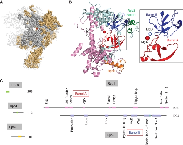
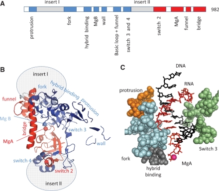
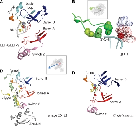
DNA transcription depends on multimeric RNA polymerases that are exceptionally conserved in all cellular organisms, with an active site region of >500 amino acids mainly harboured by their Rpb1 and Rpb2 subunits. Together with the distantly related eukaryotic RNA-dependent polymerases involved in gene silencing, they form a monophyletic family of ribonucleotide polymerases with a similarly organized active site region based on two double-Psi barrels. Recent viral and phage genome sequencing have added a surprising variety of putative nucleotide polymerases to this protein family. These proteins have highly divergent subunit composition and amino acid sequences, but always contain eight invariant amino acids forming a universally conserved catalytic site shared by all members of the two-barrel protein family. Moreover, the highly conserved 'funnel' and 'switch 2' components of the active site region are shared by all putative DNA-dependent RNA polymerases and may thus determine their capacity to transcribe double-stranded DNA templates.


Figures

Figure 1.
Figure 1.
Conserved motifs shared by the bacterial, archaeal and eukaryotic RNAPs. (A) Conserved amino acids are shown as grey (all cellular organisms) or orange spheres (archaeal RNAPs and eukaryotic RNAP I, II and III). The red dot denotes the catalytic MgA. This figure corresponds to the sequence alignments provided as Supplementary Data S1 and is based on the 1WCM PDB coordinates showing the complete twelve-subunit structure of yeast RNAP II (11), using the Pymol software (http://www.pymol.org). (B) Spatial organization of the amino acid positions common to all bacterial, archaeal and eukaryotic RNAPs (I, II and III). Experimental data are as in Figure 1A above. Rpb1, Rpb2, Rpb3/11 and Rpb6 domains are in pink, blue, green and orange, respectively. The box illustrates the Barrel A (red) and B (blue) motifs. The corresponding yeast RNAP II positions are 346–365 and 444–487 (Rpb1) and 822–844, 974–1011 and 1074–1089 (Rpb2). (C) Distribution of the corresponding motifs on the Rpb1, Rpb2, Rpb3, Rpb6 and Rpb11 subunits. Colour symbols are as in (B) above. Individual domains are named according to ref. (7), expect for part of the Rpb2 hybrid binding domain (positions 964–1028) which is referred to as the basic loop and Rpb2 funnel domains (see text for explanations).
Figure 2.
Figure 2.
Conserved motifs of the Orf6 protein. (A) Distribution of the Orf6-specific (inserts I and II) or Rpb1 (red) and Rpb2 (blue) conserved domains on the Orf6 amino acid chain, based on the sequence alignments provided as Supplementary Data S1. (B) Spatial organization in the yeast RNAP II crystal structure (upper view). This figure is based on the 1R9S PDB coordinates showing the elongating RNAP II (8) using the same colour code and same orientation as in Figure 1B above. Grey ovals denote the hypothetical insertion sites of the Orf6-specific domains. (C) Spatial organization of the conserved protrusion, fork, hybrid binding and switch 3 domains of Rpb2 on each side the RNA/DNA hybrid structure (side view). The orange, blue, grey and green spheres highlight positions 199–210 (protrusion), 466–512 (fork), 763–778 (hybrid binding) and 1106–1131 (switch 3) of yeast Rpb2, respectively. The first nine complementary bases of the RNA and template DNA strand are shown in red and blue, respectively.
Figure 3.
Figure 3.
Conserved active centre in non-canonical RNAPs. (A) Conserved amino acids in LEF-9/Rpb1 and LEF-8/Rpb2. This figure is based on the 1R9S PDB coordinates which corresponds to the ‘entry’ position incoming NTP (8). Conserved amino acids were as defined in Supplementary Data S1. The Rpb1 and Rpb2 components of the funnel domain are in orange and light blue, respectively. The box shows the entire RNAP II structure in the same spatial orientation. (B) Conserved Lef5/TFIIS C-terminal domain. Four invariant amino acids (Q285, R287, D290 and E291 in the yeast TFIIS) are space-filled (green spheres). The dotted spheres correspond to the invariant carboxylic acids of LEF-9/Rpb1 (D479, D481, D483 of Rpb1, in red) and LEF-8/Rpb2 (D837 of Rpb2, shown in blue). The spatial organization of the corresponding TFIIS, Rpb1 and Rpb2 motifs was taken from the PDB 3GTM2 coordinates which represent RNAP II in its back-tracking conformation (36). The box shows the 10-subunit RNAP II structure in the same spatial orientation. (C) Conserved amino acids in gp275, gp139 and gp273/274 (phage 201ϕ2). Conserved amino acids were as defined in Supplementary Data S1. This figure is based on the 1R9S PDB coordinates (8), with the same colour code. The bridge, trigger and Zn8/lid domains, which are additionally conserved in gp275, gp130 and gp139 (see Supplementary Data S1) are shown in green, yellow and grey, respectively. Same spatial organization as in (A). (D) Conserved amino acids in NCgl 1702 (C. glutamicum). Same spatial organization as in (A).
Figure 4.
Figure 4.
A minimal active site shared by all two-barrel RNAPs. (A) Homology of the Neurospora crassa RNA-dependent RNAPs (Qde1 and Sad1). The two sequences were aligned by a Blosum 6 matrix at a stringency of 6/23, using the Strider 1.4f6 software (61). (B) Spatial organization of the conserved domain in eukaryotic RNA-dependent RNAPs. This figure is based on the 2J7N PDB coordinates (13). Positions 691–747 (blue) and 955–1014 (red) correspond to the conserved domains shown as in Supplementary Data S2. The less conserved part of the two barrels is in dark grey. The rest of the Qde1 structure, which is poorly conserved, is represented by light grey lines. The red sphere denotes MgA. (C) Blow-up of the eight invariant amino acids shared by all two barrel RNAPs in Qde1 (N. crassa). Spheres correspond to MgA, MgB and eight invariant amino acid positions shown in red (D709, D1007, D1009 and D1011), blue (R738, K743, R962) and grey (P964), based on the PDB 2J7N coordinates (14). (D) Blow-up of the eight invariant amino acids shared by all two barrel RNAPs in RNAP II (S. cerevisiae). The corresponding invariant positions are Rpb1 D479, D481, D483 and Rpb2-D837 (red), Rpb1-R446, Rpb2-K979 and Rpb2-K987 (blue) and Rpb1-P448 (grey), as taken from PDB 1R9T (8).
Figure 5.
Figure 5.
NTP loading and organization of the switch 2 domain in yeast RNAP II. (A) NTP loading. The left and right panel correspond to the NTP entry and addition configurations, respectively, and are based on the PDB 1R9T and 1R9S coordinates (8). The invariant Rpb1-R446 (light blue), the Rpb2-K979 and Rpb2-K987 of the basic loop (dark blue), and the Rpb1-K752 and Rpb2-R1020 of the funnel domain (orange and green) are shown as space-filled spheres. The red and magenta spheres denote the catalytic MgA and MgB, respectively. The incoming NTP is shown in yellow. Thin black lines indicate the last two nucleotides of the RNA transcript. A blue arrow symbolises the rotation of the NTP toward the 3′-OH end of the transcript. (B) View of the entry site showing the surface of the RNAP II molecule at the end of the secondary channel (NTP entry pore). Same colour code as above, with additional indication of the DFDGD MgA loop. (C) Switch 2 domain: the switch 2 motif (Rpb1327–351) is shown in magenta. Space-filled spheres denote positively charged residues. Crystallographic coordinates were taken from PDB 1R9T (8).

Similar articles

Cited by

References

    1. Cermakian N, Ikeda TM, Miramontes P, Lang BF, Gray MW, Cedergren R. On the evolution of the single-subunit RNA polymerases. J. Mol. Evol. 1997;45:671–681. - PubMed
    1. Werner F, Weinzierl RO. A recombinant RNA polymerase II-like enzyme capable of promoter-specific transcription. Mol. Cell. 2002;10:635–646. - PubMed
    1. Zhang G, Campbell EA, Minakhin L, Richter C, Severinov K, Darst SA. Crystal structure of Thermus aquaticus core RNA polymerase at 3.3 A resolution. Cell. 1999;98:811–824. - PubMed
    1. Korkhin Y, Unligil UM, Littlefield O, Nelson PJ, Stuart DI, Sigler PB, Bell SD, Abrescia NG. Evolution of Complex RNA Polymerases: The Complete Archaeal RNA Polymerase Structure. PLoS Biol. 2009;7:e102. - PMC - PubMed
    1. Hirata A, Klein BJ, Murakami KS. The X-ray crystal structure of RNA polymerase from Archaea. Nature. 2008;451:851–854. - PMC - PubMed

Publication types

MeSH terms