Clinical Trial
. 2010 May;133(Pt 5):1312-27.
doi: 10.1093/brain/awq056. Epub 2010 Mar 31.

Beneficial effect of human anti-amyloid-beta active immunization on neurite morphology and tau pathology

Affiliations
Clinical Trial

Beneficial effect of human anti-amyloid-beta active immunization on neurite morphology and tau pathology

Alberto Serrano-Pozo et al. Brain. 2010 May.

Abstract

Anti-amyloid-beta immunization leads to amyloid clearance in patients with Alzheimer's disease, but the effect of vaccination on amyloid-beta-induced neuronal pathology has not been quantitatively examined. The objectives of this study were to address the effects of anti-amyloid-beta active immunization on neurite trajectories and the pathological hallmarks of Alzheimer's disease in the human hippocampus. Hippocampal sections from five patients with Alzheimer's disease enrolled in the AN1792 Phase 2a trial were compared with those from 13 non-immunized Braak-stage and age-matched patients with Alzheimer's disease, and eight age-matched non-demented controls. Analyses included neurite curvature ratio as a quantitative measure of neuritic abnormalities, amyloid and tau loads, and a quantitative characterization of plaque-associated neuritic dystrophy and astrocytosis. Amyloid load and density of dense-core plaques were decreased in the immunized group compared to non-immunized patients (P < 0.01 and P < 0.001, respectively). The curvature ratio in non-immunized patients with Alzheimer's disease was elevated compared to non-demented controls (P < 0.0001). In immunized patients, however, the curvature ratio was normalized when compared to non-immunized patients (P < 0.0001), and not different from non-demented controls. In the non-immunized patients, neurites close to dense-core plaques (within 50 microm) were more abnormal than those far from plaques (i.e. beyond 50 microm) (P < 0.0001). By contrast, in the immunized group neurites close to and far from the remaining dense-core plaques did not differ, and both were straighter compared to the non-immunized patients (P < 0.0001). Compared to non-immunized patients, dense-core plaques remaining after immunization had similar degree of astrocytosis (P = 0.6060), more embedded dystrophic neurites (P < 0.0001) and were more likely to have mitochondrial accumulation (P < 0.001). In addition, there was a significant decrease in the density of paired helical filament-1-positive neurons in the immunized group as compared to the non-immunized (P < 0.05), but not in the density of Alz50 or thioflavin-S positive tangles, suggesting a modest effect of anti-amyloid-beta immunization on tangle pathology. Clearance of amyloid plaques upon immunization with AN1792 effectively improves a morphological measure of neurite abnormality in the hippocampus. This improvement is not just attributable to the decrease in plaque load, but also occurs within the halo of the remaining dense-core plaques. However, these remaining plaques still retain some of their toxic potential. Anti-amyloid-beta immunization might also ameliorate the hippocampal tau pathology through a decrease in tau phosphorylation. These data agree with preclinical animal studies and further demonstrate that human anti-amyloid-beta immunization does not merely clear amyloid from the Alzheimer's disease brain, but reduces some of the neuronal alterations that characterize Alzheimer's disease.


Figures
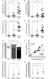
Figure 1
Figure 1
Decreased hippocampal amyloid deposition after anti-Aβ immunization. Plaque amyloid load in AN1792-treated Alzheimer’s disease patients is reduced down to the levels of non-demented controls (NC) (A), without significant increase of vascular amyloid load (B). Density of both total amyloid plaques (C) and dense-core plaques (D) is significantly decreased in the immunized group. Bars in scatter-dot plots A–D denote mean ± SEM. Two-tailed unpaired t-tests were run for these pairwise comparisons (n.s. = non-significant; **P < 0.01; ***P < 0.001; #P < 0.0001). (E, F) The proportion of dense-core plaques is significantly increased after immunization. In E, the bar graph shows total amyloid plaques counted on sections immunostained for 3D6 anti-Aβ antibody normalized to 100%. Filled bars represent the percent of amyloid plaques co-stained with thioflavin-S (dense-core plaques). Raw fractions are presented in parenthesis within the filled bars, with the number of total amyloid plaques counted in the denominator and the number of dense-core plaques in the numerator. In F, the correlation between total amyloid plaques and dense-core plaques in both Alzheimer’s disease groups is shown. Pairwise comparisons in E were done with χ2 with Fisher’s exact test (#P < 0.0001), whereas correlations in F were performed with Pearson’s test. (G, H) Remaining total amyloid plaques and dense-core plaques in immunized patients are significantly smaller compared to plaques in non-immunized patients, whereas their size does not differ between non-immunized patients and non-demented controls. Single-symbol plots in G and H represent median values, whereas bars denote interquartile ranges. These pairwise comparisons were performed with a Mann–Whitney test (**P < 0.01, #P < 0.0001). Automatic measurement of total amyloid plaques size included 225 plaques from non-demented controls (NC), 626 plaques from immunized patients (AD + TX) and 3973 plaques from non-immunized patients (AD w/o TX). Manual measurement of dense-core plaques size was performed on 55 plaques from non-demented controls, 285 plaques from immunized patients and 1035 plaques from non-immunized patients.
Figure 2
Figure 2
Improvement of neurite trajectories in immunized patients with Alzheimer’s disease. (A, B) Neurite curvature ratio correlates significantly with the density of total and dense-core amyloid plaques in non-immunized patients with Alzheimer’s disease. Correlations were performed with Spearman’s rank test. Dotted lines represent the 95% confidence intervals. (C) Neurite trajectories in the hippocampus of immunized patients are significantly straighter compared to non-immunized patients and similar to non-demented controls. Curvature ratio was analysed in 1231 neurites from 8 normal controls, 580 neurites from 4 immunized patients and 2243 neurites from 13 non-immunized patients. In one of the immunized cases an appropriate neurofilament heavy chain immunostaining was not feasible, probably due to differences in fixation protocols. (D) Improvement of curvature ratio in immunized patients is not only attributable to a lower plaque load but also occurs within the vicinity ( < 50 µm) of remaining dense-core plaques. Curvature ratio was determined in 321 neurites close to and 260 neurites far from plaques in the immunized group, and in 1256 neurites close to and 972 neurites far from plaques in the non-immunized group. Pairwise comparisons in C and D were performed with Mann–Whitney test (#P < 0.0001). (E) Curvature ratio of neurites close to plaques is not simply due to a ‘mass effect’ caused by the plaques because plaque size does not affect the curvature ratio of surrounding neurites (within-group comparisons were not significant but are not illustrated for clarity purposes). Moreover, the improvement of the curvature ratio close to plaques in the immunized subjects is not explained by the smaller size of their plaques because between-group significant difference is preserved across different plaque size intervals. This analysis included 143, 281, 202, 171 and 276 neurites close to plaques of increasing size intervals from the non-immunized Alzheimer’s disease subjects, and 112, 59, 41, 18 and 50 neurites close to plaques of the same size intervals from the AN1792-treated Alzheimer’s disease subjects. A Kruskal–Wallis ANOVA with Dunn’s multiple comparison post-test was run (***P < 0.001, #P < 0.0001, n.s. = non significant). (F, G) Representative pictures of neurite segments within the vicinity of a dense-core plaque in the CA1 region of a non-immunized patient with Alzheimer’s disease (F, Case 1), and an AN1792-treated patient with Alzheimer’s disease (G, Case 22). Neurites were immunostained with a neurofilament heavy chain antibody (in red) and plaques were stained with thioflavin-S (in green). Neurite curvature ratio was calculated as the ratio of the measured length (white line) to the end-to-end length (pink line). Compare the abnormally curve trajectory of neurites in F with the straighter trajectory of neurites in G. Scale bars = 20 µm. NC = non-demented controls; AD + TX = plaques from immunized patients; AD w/o TX = non-immunized patients.
Figure 3
Figure 3
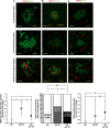
Dense-core plaques remaining after immunization retain some of their toxic properties. (A) Representative images of dense-core plaques (in green) from a non-demented control (Case 19), a non-immunized patient (Case 2) and an AN1792-treated patient (Case 23), with the neurofilament antibody SMI312 depicting associated dystrophic neurites and axonal varicosities or swellings (in red). Quantification of the number of dystrophic neurites and varicosities per plaque yielded a significant increase in the amount of neuritic dystrophy in the remaining plaques of immunized patients, as compared to non-immunized patients. Pairwise comparisons were done with the Mann–Whitney test [***P < 0.001, #P < 0.0001; n = 55, 285 and 1035 plaques in normal controls (NC), immunized patients (AD + TX) and non-immunized patients (AD w/o TX), respectively]. (B) Representative images of mitochondria accumulation in dense-core plaques associated-dystrophic neurites from a non-demented control (Case 19), a non-immunized patient (Case 3) and an AN1792-treated patient (Case 22), as revealed by the mitochondrial marker VDAC1 (in red). Quantification of dense-core plaques immunoreactive for VDAC1 in the three study groups revealed an increase of the proportion of plaques VDAC1-positive in the immunized group, as compared to the non-immunized group. Dense-core plaques were normalized to 100% and filled bars represent the proportion of dense-core plaques immunoreactive for VDAC1. Raw fractions are shown in parenthesis within the filled bars, with the number of dense-core plaques counted in the denominator and the number of VDAC1-positive plaques in the numerator. Pairwise comparisons were done with χ2 with Fisher’s exact test (*P < 0.05, ***P < 0.001). (C) Representative images of reactive astrocytosis surrounding dense-core plaques (in green) from a non-demented control (Case 18), a non-immunized patient (Case 5) and a patient treated with AN1792 (Case 23), as shown with a glial fibrillar acid protein immunostaining (in red). Quantification of the number of astrocytes per plaque resulted in a non-significant decrease of plaque-associated astrocytosis in the immunized group compared to the non-immunized patients. However, plaques from immunized patients still have more severe astrocytosis than plaques from non-demented controls. Pairwise comparisons were done with the Mann–Whitney test (***P < 0.001, #P < 0.0001; n = 66, 151 and 994 dense-core plaques, in the non-demented, immunized and non-immunized groups, respectively). Scale bars in A–C = 20 µm.
Figure 4
Figure 4
Decreased tau phosphorylation in neurofibrillary tangles after anti-Aβ immunization. (A) Hippocampal density of PHF1-positive neurons is significantly decreased in the immunized Alzheimer’s disease patients compared to the patients with non-immunized Alzheimer’s disease, despite both groups being matched for Braak stage. (B, C) No significant difference is observed in the densities of Alz50-positive neurons and thioflavin-S positive neurofibrillary tangles (NFT) between both Alzheimer’s disease groups. Pairwise comparisons in A–C were done with a two-tailed t-test and bars represent mean ± SEM (*P < 0.05, **P < 0.01, #P < 0.0001). (D) Correlations between densities of PHF1-positive neurons and Alz50-positive neurons in both Alzheimer’s disease groups reveal a predominance of the late-stage phospho-tau species (PHF1) over the early-stage misfolded tau species (Alz50) in the non-immunized group. By contrast, neither of both tau epitopes is predominant in the Braak-matched immunized Alzheimer’s disease group. Black circles represent each of non-immunized patients and grey squares represent immunized patients. Correlations were done with Pearson’s test and dotted lines indicate the 95% confidence interval. For clarity purposes, non-demented controls are not represented. (E–L) Pictures show some of the PHF1-immunostained hippocampal sections used in the above analysis. The transition from the CA1 region to the subiculum (left-to-right in each picture) is illustrated for two representative non-immunized Alzheimer’s disease patients (E, Case 4, Braak VI; and F, Case 12, Braak IV), one representative non-demented control (G, Case 18, Braak II) and the five AN1792-treated Alzheimer’s disease patients (H–L, Cases 22–26). Scale bars = 200 µm.

Comment in

Similar articles

Cited by

References

    1. Akiyama H, Tago H, Itagaki S, McGeer PL. Occurrence of diffuse amyloid deposits in the presubicular parvopyramidal layer in Alzheimer’s disease. Acta Neuropathol. 1990;79:537–44. - PubMed
    1. Augustinack JC, Schneider A, Mandelkow EM, Hyman BT. Specific tau phosphorylation sites correlate with severity of neuronal cytopathology in Alzheimer’s disease. Acta Neuropathol. 2002;103:26–35. - PubMed
    1. Bacskai BJ, Kajdasz ST, Christie RH, Carter C, Games D, Seubert P, et al. Imaging of amyloid-β deposits in brains of living mice permits direct observation of clearance of plaques with immunotherapy. Nat Med. 2001;7:369–72. - PubMed
    1. Bacskai BJ, Kajdasz ST, McLellan ME, Games D, Seubert P, Schenk D, et al. Non-Fc-mediated mechanisms are involved in clearance of amyloid-β in vivo by immunotheraphy. J Neurosci. 2002;22:7873–8. - PMC - PubMed
    1. Bard F, Cannon C, Barbour R, Burke RL, Games D, Grajeda H, et al. Peripherally administered antibodies against amyloid β-peptide enter the central nervous system and reduce pathology in a mouse model of Alzheimer disease. Nat Med. 2000;6:916–9. - PubMed

Publication types

MeSH terms