

Responsible use of computed tomography in the evaluation of coronary artery disease and chest pain
- PMID: 20360294
- PMCID: PMC2848424
- DOI: 10.4065/mcp.2009.0652
Responsible use of computed tomography in the evaluation of coronary artery disease and chest pain
Abstract
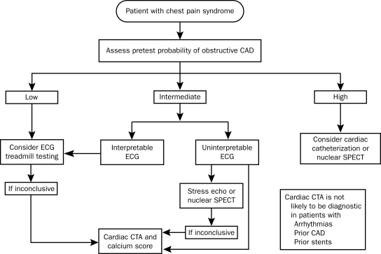
Many options are available to clinicians for the noninvasive evaluation of the cardiovascular system and patient concerns about chest discomfort. Cardiac computed tomography (CT) is a rapidly advancing field of noninvasive imaging. Computed tomography incorporates coronary artery calcium scoring, coronary angiography, ventricular functional analysis, and information about noncardiac thoracic anatomy. We searched the PubMed database and Google from inception to September 2009 for resources on the accuracy, risk, and predictive capacity of coronary artery calcium scoring and CT coronary angiography and have reviewed them herein. Cardiac CT provides diagnostic information comparable to echocardiography, nuclear myocardial perfusion imaging, positron emission tomography, and magnetic resonance imaging. A cardiac CT study can be completed in minutes. In patients with a nondiagnostic stress test result, cardiac CT can preclude the need for invasive angiography. Prognostic information portends excellent outcomes in patients with normal study results. Use of cardiac CT can reduce health care costs and length of emergency department stays for patients with chest pain. Cardiac CT examination provides clinically relevant information at a radiation dose similar to well-established technologies, such as nuclear myocardial perfusion imaging. Advances in technique can reduce radiation dose by 90%. With appropriate patient selection, cardiac CT can accurately diagnose heart disease, markedly decrease health care costs, and reliably predict clinical outcomes.
Figures
Comment in
-
Emergency department assessment of acute-onset chest pain: contemporary approaches and their consequences.Mayo Clin Proc. 2010 Apr;85(4):309-13. doi: 10.4065/mcp.2010.0141. Mayo Clin Proc. 2010. PMID: 20360290 Free PMC article. No abstract available.
References
-
- Gerber TC, Kuzo RS, Karstaedt N, et al. Current results and new developments of coronary angiography with use of contrast-enhanced computed tomography of the heart. Mayo Clin Proc. 2002;77(1):55-71 - PubMed
-
- Lanzer P, Garrett J, Lipton MJ, et al. Quantitation of regional myocardial function by cine computed tomography: pharmacologic changes in wall thickness. J Am Coll Cardiol. 1986;8(3):682-692 - PubMed
-
- Miller JC, Abbara S, Mamuya WS, Thrall JH, Uppot RN. Dual-source CT for cardiac imaging. J Am Coll Radiol. 2009;6(1):65-68 - PubMed
-
- Oncel D, Oncel G, Tastan A. Effectiveness of dual-source CT coronary angiography for the evaluation of coronary artery disease in patients with atrial fibrillation: initial experience. Radiology 2007;245(3):703-711 - PubMed
-
- Agatston AS, Janowitz WR, Hildner FJ, Zusmer NR, Viamonte M, Jr, Detrano R. Quantification of coronary artery calcium using ultrafast computed tomography. J Am Coll Cardiol. 1990;15(4):827-832 - PubMed
