

Feasibility of angiographic CT in peri-interventional diagnostic imaging: a comparative study with multidetector CT
- PMID: 20360343
- PMCID: PMC7965444
- DOI: 10.3174/ajnr.A2086
Feasibility of angiographic CT in peri-interventional diagnostic imaging: a comparative study with multidetector CT
Abstract
Background and purpose: The ability to perform neuroimaging on the angiography suite is important in making decisions during neurointerventions. Our aim was the evaluation of ACT as a fast available diagnostic tool during and after neuroendovascular procedures and the comparison of ACT with postinterventional MDCT.
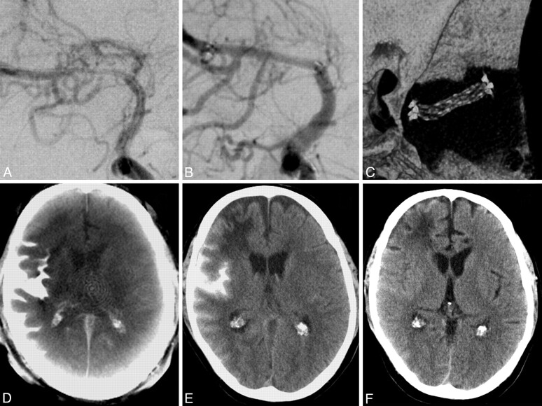
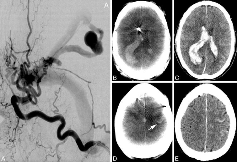
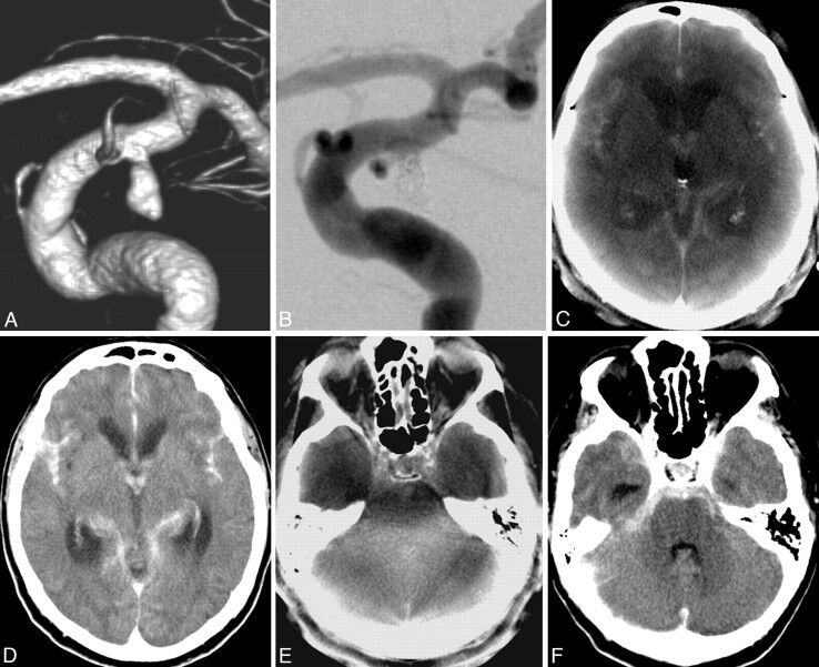
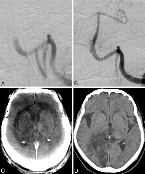
Materials and methods: Eighty-four peri-interventional ACT acquisitions were obtained and evaluated: 38 after coil embolization of cerebral aneurysms, 16 after intracranial angioplasty with stent placement, and 30 after endovascular mechanical thrombectomy and lysis. Interventions and ACTs were performed on a biplane angiography system equipped with flat panel detectors. Postprocessing was performed on a dedicated workstation, and multiplanar reformations were generated. Reference studies were performed on a 16- or 128-section MDCT scanner. All studies were independently evaluated by 3 blinded neuroradiologists. The Wilcoxon test was applied for the statistical analysis.
Results: ACT and MDCT images were of equal diagnostic quality in most cases related to the supratentorial ventricular system and the detection of hemorrhages (subarachnoidal, intraparenchymal, and intraventricular). Regarding the supratentorial ventricular system, an adequate diagnostic quality was assigned to 94% of the ACT acquisitions. For the detection of hemorrhage, no statistically significant difference was noted between ACT and MDCT. However, for the infratentorial region, ACT performed relatively poorly compared with MDCT. The diagnostic evaluation of gray matter (basal ganglia, insular cortex, and central cortex) by ACT is not sufficient, with <20% of the acquisitions scoring a diagnostic value.
Conclusions: After neuroendovascular procedures and within the angiography suite, ACT enables an immediate detection of peri-interventional hemorrhage or hydrocephalus. However, for the detection of cerebral infarction, ACT is not yet reliable.
Figures
References
-
- Doelken M, Struffert T, Richter G, et al. Flat-panel detector volumetric CT for visualization of subarachnoid hemorrhage and ventricles: preliminary results compared to conventional CT. Neuroradiology 2008;50:517–23 - PubMed
-
- Kalender WA. The use of flat-panel detectors for CT imaging [in German]. Radiologe 2003;43:379–87 - PubMed
-
- Loose R, Wucherer M, Brunner T, et al. Visualization of 3D low contrast objects by CT cone-beam reconstruction of a rotational angiography with a dynamic solid body detector [in German]. Rofo 2005;S1:PO160
-
- Groh BA, Siewerdsen JH, Drake DG, et al. A performance comparison of flat panel imager-based MV and kV cone-beam CT. Med Phys 2002;29:967–75 - PubMed
Publication types
MeSH terms
LinkOut - more resources
Full Text Sources
Medical