Review
. 2011 Feb;32(2):230-7.
doi: 10.3174/ajnr.A2073. Epub 2010 Apr 1.

Syndromes of the first and second branchial arches, part 2: syndromes

Affiliations
Review

Syndromes of the first and second branchial arches, part 2: syndromes

J M Johnson et al. AJNR Am J Neuroradiol. 2011 Feb.

Abstract

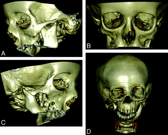
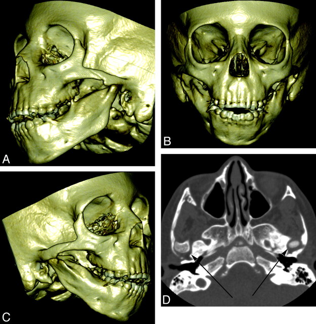
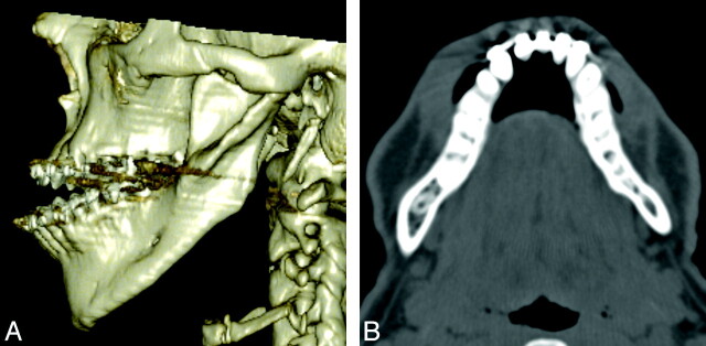
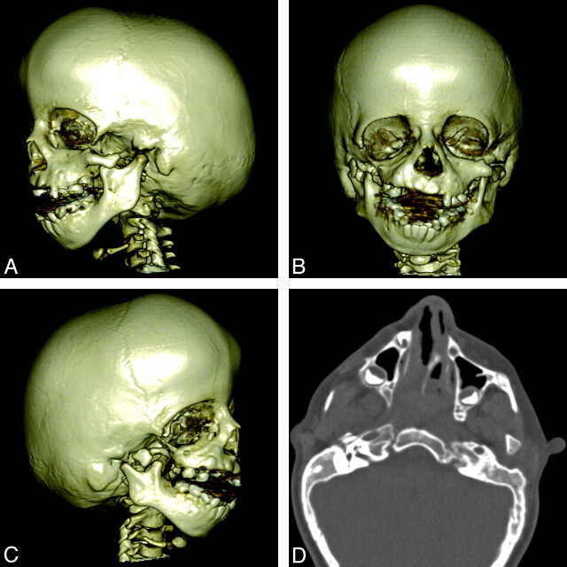
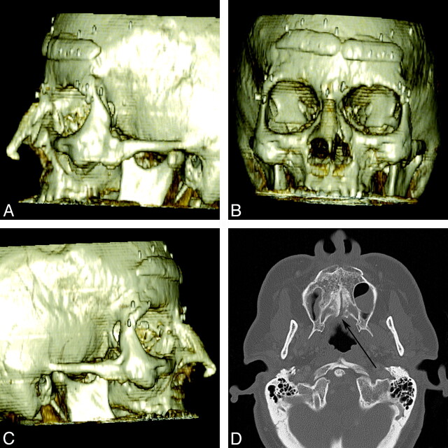
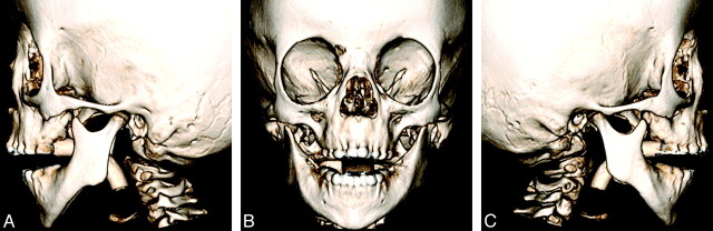
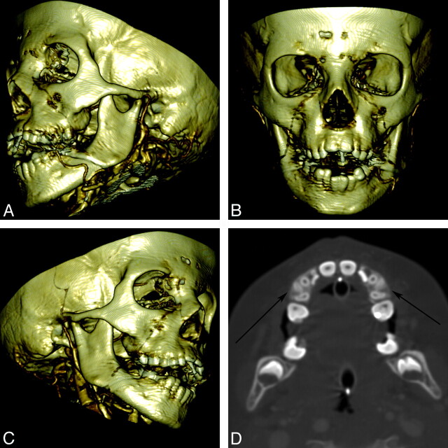
A variety of congenital syndromes affecting the face occur due to defects involving the first and second BAs. Radiographic evaluation of craniofacial deformities is necessary to define aberrant anatomy, plan surgical procedures, and evaluate the effects of craniofacial growth and surgical reconstructions. High-resolution CT has proved vital in determining the nature and extent of these syndromes. The radiologic evaluation of syndromes of the first and second BA should begin first by studying a series of isolated defects (cleft lip with or without CP, micrognathia, and EAC atresia) that compose the major features of these syndromes and allow a more specific diagnosis. After discussion of these defects and the associated embryology, we discuss PRS, HFM, ACS, TCS, Stickler syndrome, and VCFS.


Figures

Fig 1.
Fig 1.
Young adult with hemifacial microstomia. A−C, 3D bone reconstruction shows right mandibular and maxillary hypoplasia compared with the normal-appearing left condyle. D, A 3-year-old boy with hemifacial microstomia. 3D bone reconstruction shows a more dramatic appearance of asymmetric hypoplasia of the mandible.
Fig 2.
Fig 2.
A 5-year-old boy with ACS. A−C, 3D bony reconstructions show an absent condylar processes, asymmetric micrognathia, and hypoplastic condyles. D, Axial CT scan shows bilateral abnormal TMJs (arrows) with dysplastic condylar processes.
Fig 3.
Fig 3.
An 18-year-old man with ACS. A, 3D bony reconstruction shows micrognathia with overprotrusion of the mandible in relation to the maxilla. The lateral mandibular cleft is a feature commonly seen in ACS (see also Fig 2). B, Axial CT scan also shows the asymmetry of the dysplastic protruding mandible.
Fig 4.
Fig 4.
A 4-year-old girl with TCS. A−C, 3D bone reconstructions show bilateral and asymmetric abnormalities of the mandibular condyle and coronoid process as well as maxillary hypoplasia and micrognathia. The EACs are absent bilaterally. D, Axial CT image shows bilateral hypoplastic zygomatic arches, maxillary bone dysplasia, and temporal bone abnormalities.
Fig 5.
Fig 5.
A 47-year-old woman with Stickler syndrome. A−C, 3D bony reconstructions show a flat midface with a depressed nasal bridge, short nose, anteverted nares, and zygomatic hypoplasia. Reconstructive and cosmetic hardware and implants are present. D, Axial CT scan shows incomplete fusion of the palatal bones with a posterior submucosal cleft (arrow).
Fig 6.
Fig 6.
A 6-year-old boy with Stickler syndrome. 3D bony reconstructions show the characteristic flat midface with a depressed nasal bridge. The degree of zygomatic hypoplasia is more subtle. There is mild micrognathia. An endotracheal tube is present. Courtesy of Michael Cunningham.
Fig 7.
Fig 7.
A 6-year-old boy with VCFS. A−C, 3D bone reconstructions show mild micrognathia with normal condyle and coronoid process morphology. D, Axial CT angiogram shows relatively symmetric micrognathia and areas of malocclusion (arrows).

Similar articles

Cited by

References

    1. Marsh JL. Comprehensive Care for Craniofacial Deformities. St. Louis, Missouri: Mosby; 1985
    1. Robin P. La chute de la base de la langue consideree comme une nouvelle cause de gene dans la respiration nasopharyngienne. Bull Acad de Med 1923;89:37–41
    1. Robin P. La Glossoptose: Un Grave Danger Pour Nos Enfants. Paris, France: Gaston Doin; 1929
    1. Robin P. Glossoptosis due to atresia and hypotrophy of the mandible. Am J Dis Child 1934;48:541
    1. Figueroa AA, Glupker TJ, Fitz MG, et al. . Mandible, tongue, and airway in Pierre Robin sequence: a longitudinal cephalometric study. Cleft Palate Craniofac J 1991;28:425–34 - PubMed

MeSH terms

Supplementary concepts