Review
. 2010 Apr 1;24(7):625-35.
doi: 10.1101/gad.1907710.

Understanding neuronal connectivity through the post-transcriptional toolkit

Affiliations
Review

Understanding neuronal connectivity through the post-transcriptional toolkit

Carlos M Loya et al. Genes Dev. .

Abstract

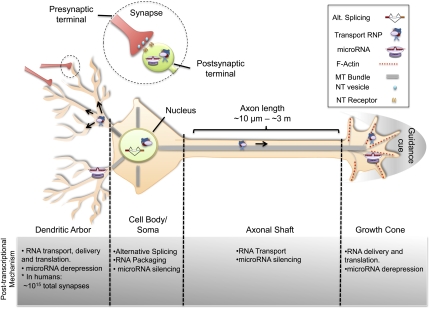
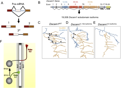
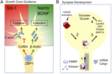
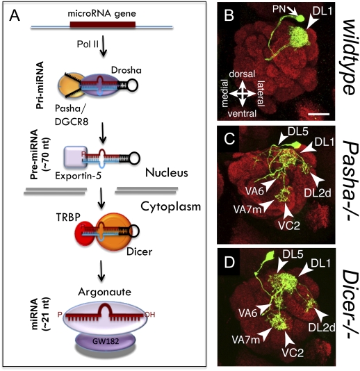
Post-transcriptional regulatory mechanisms have emerged as a critical component underlying the diversification and spatiotemporal control of the proteome during the establishment of precise neuronal connectivity. These mechanisms have been shown to be important for virtually all stages of assembling a neural network, from neurite guidance, branching, and growth to synapse morphogenesis and function. From the moment a gene is transcribed, it undergoes a series of post-transcriptional regulatory modifications in the nucleus and cytoplasm until its final deployment as a functional protein. Initially, a message is subjected to extensive structural regulation through alternative splicing, which is capable of greatly expanding the protein repertoire by generating, in some cases, thousands of functionally distinct isoforms from a single gene locus. Then, RNA packaging into neuronal transport granules and recognition by RNA-binding proteins and/or microRNAs is capable of restricting protein synthesis to selective locations and under specific input conditions. This ability of the post-transcriptional apparatus to expand the informational content of a cell and control the deployment of proteins in both spatial and temporal dimensions is a feature well adapted for the extreme morphological properties of neural cells. In this review, we describe recent advances in understanding how post-transcriptional regulatory mechanisms refine the proteomic complexity required for the assembly of intricate and specific neural networks.


Figures

Figure 1.
Figure 1.
Complexity of neuronal morphology and post-transcriptional regulation. Typical neuron illustrating four gross neuronal anatomical segments: cell body/soma, axonal shaft, dendritic arbor, and growth cone. Inset shows a synapse, with presynaptic and post-synaptic terminals. The post-transcriptional mechanisms highlighted here are AS, RNA transport, and miRNA silencing. AS occurs in the nucleus of the cell body, and is important for generating protein isoforms. Following splicing and mRNA maturation, the transcript can be packaged into transport RNPs, repressed by RBPs and/or miRNAs, delivered to distal sites of the neuron, and locally translated in response to stimulus. Moreover, miRNAs are involved in finely regulating protein levels throughout the cell.
Figure 2.
Figure 2.
Roles of AS in neuronal connectivity. (A) Forms of AS. Most eukaryotic genes consist of protein-coding exon (rectangles) and noncoding intron (lines) sequences. Following transcription, the splicesome and splicing factors mediate intron removal from pre-mRNA to create a mature mRNA transcript. AS is a mechanism by which exons are included or excluded from the mRNA (1,2), thereby creating isoforms (also called variants) of distinct protein structures. In some instances, AS may result in differential intron retention (3*), such as in the case of Robo3 (Chen et al. 2008), which generates a premature stop codon that vastly changes the C-terminal of the encoded gene. (B) Gene structure of Dscam1 ectodomains illustrating the potential AS isoforms. (C–E) Representative illustrations of dendritic arborization neurons in different Dscam1 mutant backgrounds (arrows represent dendritic branch overlap). (C) Dendrites of different dendritic arborization classes overlap extensively. (D) However, in Dscam1 mutants expressing 1152 isoforms, the amount of overlap between class I and III dendrites decreased significantly. (E) When Dscam1 diversity decreased to 12 or 24, dendrites of distinct classes completely avoid each other, indicating that appropriate patterning of dendritic arborization neurons depends on thousands of Dscam1 isoforms (Hattori et al. 2009). (F) Schematic of a commissural neuron during embryonic development as seen in an open book spinal cord dissection. Axonal shafts represent the differential expression patterns of Robo3 isoforms. In this context, Robo3.1 enables axons to cross the midline, whereas Robo3.2 prevents axons from recrossing (Chen et al. 2008).
Figure 3.
Figure 3.
RNA transport and local translation. (A) Illustration of a growth cone exposed to chemoattractant (Netrin and BDNF) and chemorepulsive (Slit-1) cues. ZBP1 regulates the delivery and local translation of β-Actin and cofilin mRNA during guidance cue-induced growth cone extension or collapse (Leung et al. 2006; Piper et al. 2006; Yao et al. 2006). (B) Schematic of a dendritic spine representing the role of FMRP in synapse morphogenesis. FMRP is involved in the kinesin-dependent transport of mRNA to synaptic sites (1). Activation of metabotropic glutamate receptors (mGluRs) derepresses the FMRP mRNA cargo (2), induces local translation (3), and promotes dendritic spine growth (4) (Dictenberg et al. 2008).
Figure 4.
Figure 4.
miRNA regulation of neuronal connectivity. (A) miRNA biogenesis pathway. miRNA are regularly transcribed by RNA Pol II, then the primary miRNA (pri-miRNA) is processed by the RNase III endonuclease Drosha and the RBP Pasha/DGRC8 into the ∼70-nt pre-miRNA. Exportin-5 exports the pre-miRNA to the cytoplasm, where it is cleaved by the another RNase III endonuclease (Dicer) and the RBP TRBP/loquacious into the ∼21 duplex miRNA. This duplex is loaded into the argonaute- and GW182-containing RISC, where it then recognizes target mRNA and promotes destabilization or translational repression. (B–D) Representative images of Drosophila single-cell MARCM clones of olfactory PNs targeting glomeruli in the antennal lobe; images reprinted from Berdnik et al. (2008), © 2008, with permission from Elsevier. (B) In a wild-type background, anterodorsal PNs (adPNs) extend dendrites and specifically target the DL1 glomerulus. In Pasha (C) and Dicer (D) mutants, adPN innervation of DL1 is reduced, and mistargeting of DL5, DL2d, VA6, VA7m, and VC2 glomeruli is observed (Berdnik et al. 2008).

References

    1. Antar LN, Li C, Zhang H, Carroll RC, Bassell GJ. Local functions for FMRP in axon growth cone motility and activity-dependent regulation of filopodia and spine synapses. Mol Cell Neurosci. 2006;32:37–48. - PubMed
    1. Aschrafi A, Schwechter AD, Mameza MG, Natera-Naranjo O, Gioio AE, Kaplan BB. MicroRNA-338 regulates local cytochrome c oxidase IV mRNA levels and oxidative phosphorylation in the axons of sympathetic neurons. J Neurosci. 2008;28:12581–12590. - PMC - PubMed
    1. Barbee SA, Estes PS, Cziko AM, Hillebrand J, Luedeman RA, Coller JM, Johnson N, Howlett IC, Geng C, Ueda R, et al. Staufen- and FMRP-containing neuronal RNPs are structurally and functionally related to somatic P bodies. Neuron. 2006;52:997–1009. - PMC - PubMed
    1. Bartel DP, Chen CZ. Micromanagers of gene expression: The potentially widespread influence of metazoan microRNAs. Nat Rev Genet. 2004;5:396–400. - PubMed
    1. Bassell GJ, Warren ST. Fragile X syndrome: Loss of local mRNA regulation alters synaptic development and function. Neuron. 2008;60:201–214. - PMC - PubMed

Publication types

LinkOut - more resources