. 2010 Apr 1;24(7):696-707.
doi: 10.1101/gad.1859310.

The nuclear hormone receptor Coup-TFII is required for the initiation and early maintenance of Prox1 expression in lymphatic endothelial cells

Affiliations

The nuclear hormone receptor Coup-TFII is required for the initiation and early maintenance of Prox1 expression in lymphatic endothelial cells

R Sathish Srinivasan et al. Genes Dev. .

Abstract

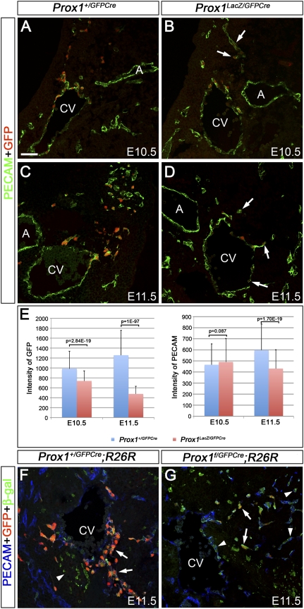
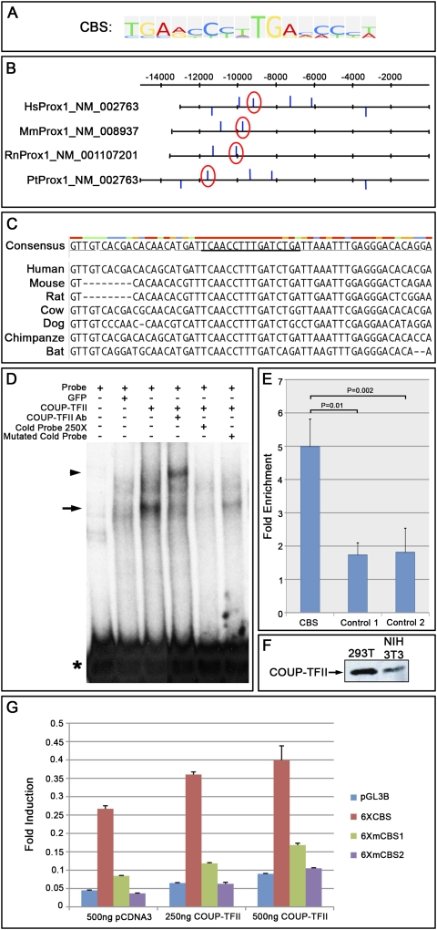
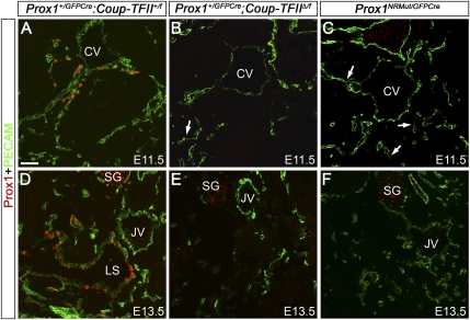
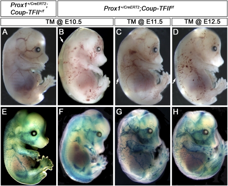
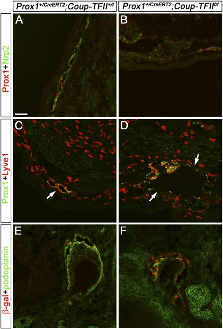
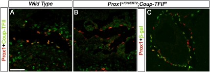
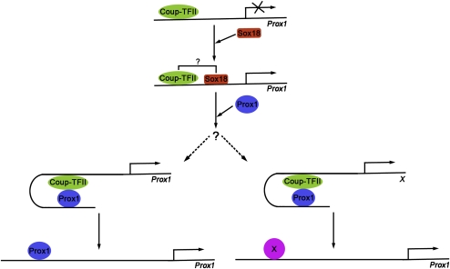
The homeobox gene Prox1 is crucial for mammalian lymphatic vascular development. In the absence of Prox1, lymphatic endothelial cells (LECs) are not specified. The maintenance of LEC identity also requires the constant expression of Prox1. However, the mechanisms controlling the expression of this gene in LECs remain poorly understood. The SRY-related gene Sox18 is required to induce Prox1 expression in venous LEC progenitors. Although Sox18 is also expressed in embryonic arteries, these vessels do not express Prox1, nor do they give rise to LECs. This finding suggests that some venous endothelial cell-specific factor is required for the activation of Prox1. Here we demonstrate that the nuclear hormone receptor Coup-TFII is necessary for the activation of Prox1 in embryonic veins by directly binding a conserved DNA domain in the regulatory region of Prox1. In addition, we show that the direct interaction between nuclear hormone receptors and Prox1 is also necessary for the maintenance of Prox1 expression during early stages of LEC specification and differentiation.


Figures

Figure 1.
Figure 1.
Maintenance of Prox1 expression in LECs requires Prox1. (A,B) Compared with that in E10.5 Prox1+/GFPCre embryos (A), Prox1 expression in Prox1LacZ/GFPCre embryos (B, arrows) is moderately reduced in the ECs migrating from the anterior cardinal vein (CV), as indicated by immunostaining for GFP (red). (C,D) At E11.5, the level of GFP expression remains high in the LECs of Prox1+/GFPCre embryos (C), whereas it is down-regulated in the Prox1-null ECs of Prox1LacZ/GFPCre littermates (D, arrows). (E) Image analysis was performed on GFP+ cells from eight identical level sections for each embryo. At E10.5, quantification of GFP and PECAM intensities indicates a moderate reduction in GFP expression in the ECs of Prox1LacZ/GFPCre embryos; PECAM levels appear comparable. At E11.5, the GFP level is markedly reduced in Prox1LacZ/GFPCre ECs. PECAM expression is only moderately reduced in Prox1-null ECs. (F) At E11.5, GFP+PECAM+β-gal+ cells line the cardinal vein and migrate from it in Prox1+/GFPCre;R26R embryos (arrows). A few mesenchymal cells weakly express GFP. Correspondingly, these cells are also β-gal+ (arrowhead). (G) GFP expression is weak in numerous β-gal+PECAM+ cells of E11.5 Prox1f/GFPCre;R26R embryos (arrows). Numerous GFPPECAM+β-gal+ cells are also located on and outside the cardinal vein (arrowhead). The neural tube is oriented toward the top of A–D and toward the right side in F and G. (CV) Anterior cardinal vein; (LS) lymph sacs; (A) dorsal aorta. Bar, 50 μm.
Figure 2.
Figure 2.
Coup-TFII binds directly to a conserved site present in the Prox1 upstream regulatory region. (A) Coup-TFII recognizes a 16-nt motif (CBS) that consists of two similar 8-nt repeats. (B) A DNA region located 5 kb upstream of the noncoding exon 1 and the entire intron 1 (upstream of the ATG translational start codon) of human (Hs), mouse (Mm), rat (Rn), and chimpanzee (Pt) Prox1 was analyzed, and putative CBSs were identified (blue bars). Upward bars indicate those in the sense orientation, and downward bars indicate those in the antisense orientation. Red circles highlight the CBS that is conserved among all species tested. (C) The identified CBS is also conserved among other mammals whose DNA sequences are available. The consensus DNA sequence is underlined. The colors above the nucleotides indicate the consensus strength, with red being the strongest and blue being the weakest. (D) The conserved CBS from the mouse Prox1 gene was amplified by PCR, and the radiolabeled probe was generated using 32P-dCTP. EMSA was performed using 293T cell lysates with or without ectopically expressed COUP-TFII. (Lane 1) Probe alone (asterisk). (Lane 2) Labeled probe incubated with GFP-transfected 293T cell lysate. (Lane 3) Labeled probe incubated with COUP-TFII-transfected 293T cell lysate. The shift in the mobility of the probe is seen (arrow). (Lane 4) Probe incubated with COUP-TFII-transfected 293T cell lysate and a mouse monoclonal antibody against COUP-TFII. A supershifted band (arrowhead) can be seen. (Lane 5) An excess (250-fold) of nonradiolabeled probe efficiently competed with the binding of COUP-TFII to the radiolabeled probe. (Lane 6) Replacement of the two highly conserved TG residues by AA residues in the nonradiolabeled probe reduced this competition. (E) ChIP was performed on human LECs maintained in culture by using a rabbit polyclonal antibody against COUP-TFII. Real-time PCR was carried out using the pulled-down DNA fragment as a template and primers and probes specific for the conserved CBS, a nonconserved CBS (control 1), or a nonspecific site 40 kb downstream from the ATG (control 2). When compared with controls, a statistically significant (P ≤ 0.01) enrichment was observed for the conserved CBS. (F) Western blotting shows the expression of COUP-TFII in 293T and NIH 3T3 cells commonly used for luciferase assays. (G) Dual luciferase assay was carried out using the generated reporters containing six consecutive conserved CBSs that are wild-type (6XCBS), carrying a mutation in 4 of the 16 nt (6XmCBS1), or carrying a mutation in 8 of the 16 nt (6XmCBS2). The 6XCBS showed endogenous activity caused by the presence of COUP-TFII in the cells. This activity increased with increasing concentrations of the COUP-TFII expression plasmid. Although the 6XmCBS1 showed reduced endogenous activity, it moderately responded to ectopic COUP-TFII. The 6XmCBS2 did not show any endogenous activity and did not respond to COUP-TFII.
Figure 3.
Figure 3.
Interaction between Coup-TFII and Prox1 is required to maintain Prox1 expression in LECs. (A) At E11.5, Prox1-expressing LECs (red) are seen in and around the anterior cardinal vein (CV) of Prox1+/GFPCre;Coup-TFII+/f embryos. (B) Just a few Prox1+ LECs (arrow) are seen in Prox1+/GFPCre;Coup-TFIIΔ/f littermates. (C) E11.5 Prox1NRMut/GFPCre embryos expressing a form of Prox1 mutated in the nuclear hormone receptor-binding site also have a reduced number of LECs (arrows). (D) At E13.5, the lymph sacs (LS) lined by Prox1+ LECs are seen in control embryos. However, at this stage, no LECs are seen in Prox1+/GFPCre;Coup-TFIIΔ/f (E) or Prox1NRMut/GFPCre (F) embryos. PECAM is shown in green. The neural tube is oriented toward the left side of all panels. (JV) Jugular vein; (SG) sympathetic ganglia. Bar, 50 μm.
Figure 4.
Figure 4.
Coup-TFII has a time-dependent role in the regulation of lymphatic vascular development. (A–D) Prox1+/CreERT2;Coup-TFII+/f (A) and Prox1+/CreERT2;Coup-TFIIf/f (B–D) embryos were exposed to TM at the indicated time points and isolated at E15.5. Blood-filled superficial vessels and edema (arrows) are observed in Prox1+/CreERT2;Coup-TFIIf/f embryos. (E–H) The above embryos were whole-mount X-gal-stained for β-gal activity. (E) The X-gal+ LECs are seen forming the dermal lymphatic plexus in control embryos. (F) In contrast, in Prox1+/CreERT2;Coup-TFIIf/f embryos exposed to TM at E10.5, the superficial lymphatic plexus is nearly absent. (G,H) The number of superficial X-gal+ cells increases concomitantly with the later time of TM exposure. However, the overall size of the lymphatic plexus remains smaller than that of control embryos.
Figure 5.
Figure 5.
Conditional deletion of CoupTFII using Prox1+/CreERT2 reduces the expression of LEC markers. Prox1+/CreERT2;CoupTFII+/f and Prox1+/CreERT2;CoupTFIIf/f embryos were exposed to 5 mg TM at E12.5 and were analyzed at E15.5 by immunostaining of sections with different LEC markers. (A) Prox1 (red) and Nrp2 (green) are coexpressed in the LECs of a peripheral lymphatic vessel of Prox1+/CreERT2;CoupTFII+/f embryos. (B) However, in Prox1+/CreERT2;CoupTFIIf/f embryos, these vessels are dilated and mispatterned, and the expression of Nrp2 is substantially reduced. (C) Prox1 (green) and Lyve1 (red) are coexpressed in the peripheral lymphatic vessel (arrow) of Prox1+/CreERT2;CoupTFII+/f embryos. (D) In contrast, the lymphatic vessels of Prox1+/CreERT2;CoupTFIIf/f embryos are dilated, and Lyve1 expression is down-regulated in the Prox1+ LECs of the peripheral lymphatic vessels (arrows). The scattered Prox1Lyve1+ cells are macrophages. (E,F) Deletion of the floxed Coup-TFII allele results in the activation of the LacZ reporter gene that expresses β-gal (red). (E) Costaining with the LEC marker podoplanin (green) shows that these markers are coexpressed in the lymphatic vessels of Prox1+/CreERT2;CoupTFII+/f embryos. (F) In contrast, the expression of podoplanin is substantially reduced in the lymphatic vessels of Prox1+/CreERT2;CoupTFIIf/f embryos. The neural tube is oriented toward the bottom of A, and toward the right side of B–F. Bar, 50 μm.
Figure 6.
Figure 6.
Prox1 expression is normal in the remaining Coup-TFII-null LECs of E15.5 Prox1+/CreERT2;Coup-TFIIf/f embryos exposed to TM at E12.5. (A) At E15.5, dermal lymphatic vessels are lined by Prox1+Coup-TFII+ LECs in wild-type embryos. (B) The Prox1+ LECs of Prox1+/CreERT2;Coup-TFIIf/f littermates are negative for Coup-TFII. (C) β-gal staining shows the efficient deletion of Coup-TFII in the remaining Prox1-expressing LECs. The neural tube is oriented toward the right side of all panels. Bar, 50 μm.
Figure 7.
Figure 7.
Model of the roles of Coup-TFII in the regulation of Prox1 expression during lymphatic vascular development. Coup-TFII alone is not sufficient to induce Prox1 expression in LECs. Therefore, in this working model, we suggest that Coup-TFII cooperates with Sox18 to initiate Prox1 expression in LEC progenitors. Once initiated, two mechanisms may maintain Prox1 expression in differentiating LECs. Prox1 could be recruited to its own promoter due to its interaction with Coup-TFII and subsequently regulate and maintain its own expression. Alternatively, the Coup-TFII–Prox1 complex might activate an as-yet-unknown transcription factor X, which in turn maintains Prox1 expression.

References

    1. Bull LN, Roche E, Song EJ, Pedersen J, Knisely AS, van Der Hagen CB, Eiklid K, Aagenaes O, Freimer NB. Mapping of the locus for cholestasis-lymphedema syndrome (Aagenaes syndrome) to a 6.6-cM interval on chromosome 15q. Am J Hum Genet. 2000;67:994–999. - PMC - PubMed
    1. de Boer J, Williams A, Skavdis G, Harker N, Coles M, Tolaini M, Norton T, Williams K, Roderick K, Potocnik AJ, et al. Transgenic mice with hematopoietic and lymphoid specific expression of Cre. Eur J Immunol. 2003;33:314–325. - PubMed
    1. Delorme B, Dahl E, Jarry-Guichard T, Briand JP, Willecke K, Gros D, Theveniau-Ruissy M. Expression pattern of connexin gene products at the early developmental stages of the mouse cardiovascular system. Circ Res. 1997;81:423–437. - PubMed
    1. Francois M, Caprini A, Hosking B, Orsenigo F, Wilhelm D, Browne C, Paavonen K, Karnezis T, Shayan R, Downes M, et al. Sox18 induces development of the lymphatic vasculature in mice. Nature. 2008;456:643–647. - PubMed
    1. Fruhwirth M, Janecke AR, Muller T, Carlton VE, Kronenberg F, Offner F, Knisely AS, Geleff S, Song EJ, Simma B, et al. Evidence for genetic heterogeneity in lymphedema-cholestasis syndrome. J Pediatr. 2003;142:441–447. - PubMed

Publication types

Substances