

The innervation and organization of motor units in a series-fibered human muscle: the brachioradialis
- PMID: 20360433
- PMCID: PMC2886675
- DOI: 10.1152/japplphysiol.01163.2009
The innervation and organization of motor units in a series-fibered human muscle: the brachioradialis
Abstract
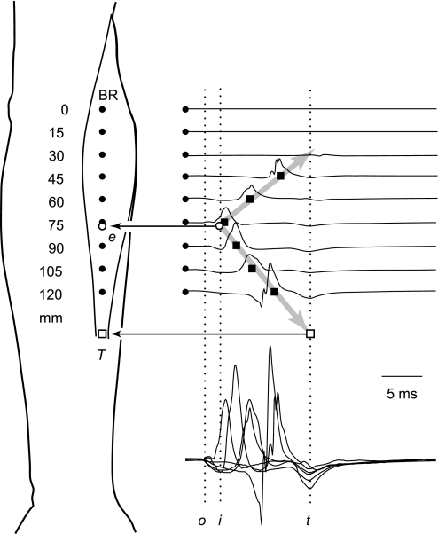
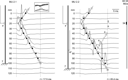
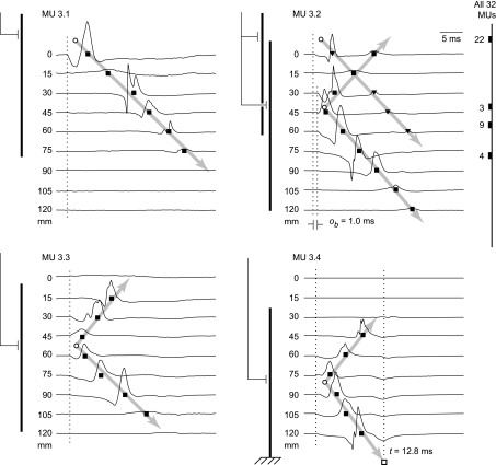
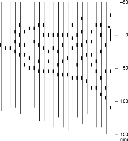
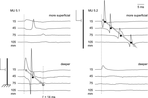
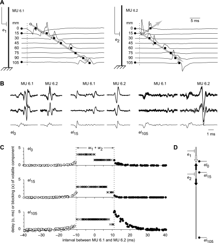
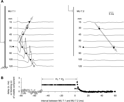
We studied the innervation and organization of motor units in the brachioradialis muscle of 25 normal human subjects. We recorded intramuscular EMG signals at points separated by 15 mm along the proximodistal muscle axis during moderate isometric contractions, identified from 27 to 61 (mean 39) individual motor units per subject using EMG decomposition, and estimated the locations of the endplates and distal muscle/tendon junctions from the motor-unit action potential (MUAP) propagation patterns and terminal standing waves. In three subjects all the motor units were innervated in a single endplate zone. In the other 22 subjects, the motor units were innervated in 3-6 (mean 4) distinct endplate zones separated by 15-55 mm along the proximodistal axis. One-third of the motor units had fibers innervated in more than one zone. The more distally innervated motor units had distinct terminal waves indicating tendonous termination, while the more proximal motor units lacked terminal waves, indicating intrafascicular termination. Analysis of blocked MUAP components revealed that 19% of the motor units had at least one doubly innervated fiber, i.e., a fiber innervated in two different endplate zones by two different motoneurons, and thus belonging to two different motor units. These results are consistent with the brachioradialis muscle having a series-fibered architecture consisting of multiple, overlapping bands of muscle fibers in most individuals and a simple parallel-fibered architecture in some individuals.
Figures
Similar articles
-
Electrophysiological evidence of doubly innervated branched muscle fibers in the human brachioradialis muscle.Clin Neurophysiol. 2007 Dec;118(12):2612-9. doi: 10.1016/j.clinph.2007.09.058. Epub 2007 Oct 31. Clin Neurophysiol. 2007. PMID: 17977064 Free PMC article.
-
Electrophysiological evidence of adult human skeletal muscle fibres with multiple endplates and polyneuronal innervation.J Physiol. 2002 Oct 15;544(2):549-65. doi: 10.1113/jphysiol.2002.023267. J Physiol. 2002. PMID: 12381826 Free PMC article.
-
Increased jitter and blocking in normal muscles due to doubly innervated muscle fibers.Muscle Nerve. 2003 Oct;28(4):423-31. doi: 10.1002/mus.10459. Muscle Nerve. 2003. PMID: 14506713
-
1998 ISEK Congress Keynote Lecture: Motor units: how many, how large, what kind? International Society of Electrophysiology and Kinesiology.J Electromyogr Kinesiol. 1998 Dec;8(6):391-402. doi: 10.1016/s1050-6411(98)00020-0. J Electromyogr Kinesiol. 1998. PMID: 9840894 Review.
-
The resilience of the size principle in the organization of motor unit properties in normal and reinnervated adult skeletal muscles.Can J Physiol Pharmacol. 2004 Aug-Sep;82(8-9):645-61. doi: 10.1139/y04-081. Can J Physiol Pharmacol. 2004. PMID: 15523522 Review.
Cited by
-
Neurophysiological Factors Affecting Muscle Innervation Zone Estimation Using Surface EMG: A Simulation Study.Biosensors (Basel). 2021 Sep 27;11(10):356. doi: 10.3390/bios11100356. Biosensors (Basel). 2021. PMID: 34677312 Free PMC article.
-
Innervation zones of fasciculating motor units: observations by a linear electrode array.Front Hum Neurosci. 2015 May 12;9:239. doi: 10.3389/fnhum.2015.00239. eCollection 2015. Front Hum Neurosci. 2015. PMID: 26029076 Free PMC article.
-
Why Temporal Inference Stimulation May Fail in the Human Brain: A Pilot Research Study.Biomedicines. 2023 Jun 24;11(7):1813. doi: 10.3390/biomedicines11071813. Biomedicines. 2023. PMID: 37509455 Free PMC article.
-
Muscle innervation zone estimation from monopolar high-density M-waves using principal component analysis and radon transform.Front Physiol. 2023 Mar 15;14:1137146. doi: 10.3389/fphys.2023.1137146. eCollection 2023. Front Physiol. 2023. PMID: 37008017 Free PMC article.
-
Detection of Multiple Innervation Zones from Multi-Channel Surface EMG Recordings with Low Signal-to-Noise Ratio Using Graph-Cut Segmentation.PLoS One. 2016 Dec 15;11(12):e0167954. doi: 10.1371/journal.pone.0167954. eCollection 2016. PLoS One. 2016. PMID: 27978535 Free PMC article.
References
-
- Abrams RA, Ziets RJ, Lieber RL, Botte MJ. Anatomy of the radial nerve motor branches in the forearm. J Hand Surg [Am] 22: 232–237, 1997 - PubMed
-
- Christensen E. Topography of terminal motor innervation in striated muscles of stillborn infants. Am J Phys Med 38: 65–78, 1959 - PubMed
-
- Duxson MJ, Sheard PW. Formation of new myotubes occurs exclusively at the multiple innervation zones of an embryonic large muscle. Dev Dyn 204: 391–405, 1995 - PubMed
-
- Eccles JC, Sherrington CS. Numbers and contraction-values of individual motor units examined in some muscles of the limb. Proc R Soc 106B: 326–357, 1930
-
- Falla D, Dall'Alba P, Rainoldi A, Merletti R, Jull G. Location of innervation zones of sternocleidomastoid and scalene muscles-a basis for clinical and research electromyography applications. Clin Neurophysiol 113: 57–63, 2002 - PubMed
Publication types
MeSH terms
Grants and funding
LinkOut - more resources
Full Text Sources