. 2010 Jun 10;115(23):4707-14.
doi: 10.1182/blood-2009-10-248872. Epub 2010 Apr 1.

Deletion of proapoptotic Puma selectively protects hematopoietic stem and progenitor cells against high-dose radiation

Affiliations

Deletion of proapoptotic Puma selectively protects hematopoietic stem and progenitor cells against high-dose radiation

Lijian Shao et al. Blood. .

Abstract

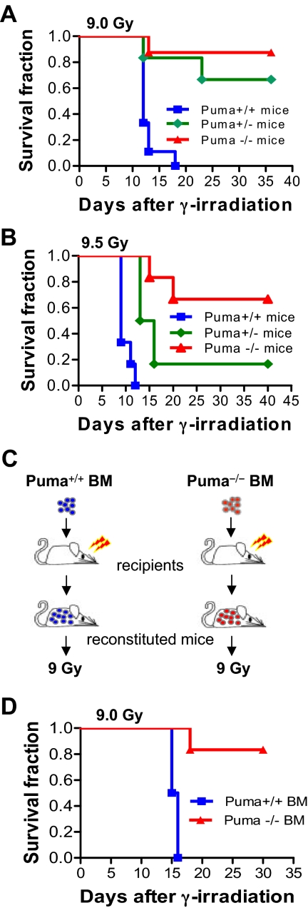
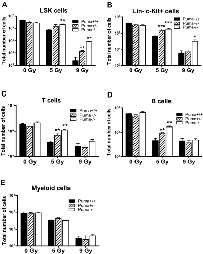
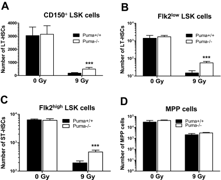
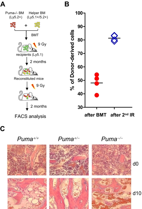
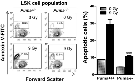
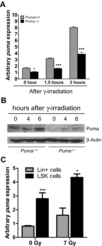
Bone marrow injury is a major adverse side effect of radiation and chemotherapy. Attempts to limit such damage are warranted, but their success requires a better understanding of how radiation and anticancer drugs harm the bone marrow. Here, we report one pivotal role of the BH3-only protein Puma in the radiosensitivity of hematopoietic stem cells (HSCs) and hematopoietic progenitor cells (HPCs). Puma deficiency in mice confers resistance to high-dose radiation in a hematopoietic cell-autonomous manner. Unexpectedly, loss of one Puma allele is sufficient to confer mice radioresistance. Interestingly, null mutation in Puma protects both primitive and differentiated hematopoietic cells from damage caused by low-dose radiation but selectively protects HSCs and HPCs against high-dose radiation, thereby accelerating hematopoietic regeneration. Consistent with these findings, Puma is required for radiation-induced apoptosis in HSCs and HPCs, and Puma is selectively induced by irradiation in primitive hematopoietic cells, and this induction is impaired in Puma-heterozygous cells. Together, our data indicate that selective targeting of p53 downstream apoptotic targets may represent a novel strategy to protecting HSCs and HPCs in patients undergoing intensive cancer radiotherapy and chemotherapy.


Figures

Figure 1
Figure 1
Deletion of Puma renders mice resistant to γ-irradiation. (A) Kaplan-Meier survival curves of mice exposed to 9 Gy TBI. Puma+/+ (n = 9), Puma+/− (n = 6), and Puma−/− (n = 8) mice were given a single dose of TBI (9 Gy) and were monitored for survival. The enhanced survival rate of Puma−/− and Puma+/− mice was significantly greater than Puma+/+ mice (P < .001 and P < .01, respectively), but the survival rate was not significantly different between Puma−/− mice versus Puma+/− mice (P > .05). (B) Kaplan-Meier survival curves of mice exposed to 9.5 Gy TBI. Puma+/+ (n = 6), Puma+/− (n = 9), and Puma−/− (n = 7) mice were then administrated a lethal TBI dose (9.5 Gy) and monitored daily for survival. The survival rate of Puma−/− and Puma+/− mice was significantly greater than Puma+/+ mice (Puma−/− mice vs Puma+/+ mice, P < .001; Puma+/− mice vs Puma+/+ mice, P < .01). (C) Diagram for generation of reconstituted mice. C57BL6/N recipients were lethally irradiated for a total of 13 Gy (6.5 Gy, 2 times, 3 hours apart) and received 1 × 10⁷ total BM cells from Puma+/+ mice or Puma−/− mice (n = 6 mice/group). (D) Kaplan-Meier survival curves of reconstituted mice after 9 Gy TBI. After 8 weeks, the mice in panel C were given a second course of TBI (9 Gy) and monitored for survival. The survival rate of mice reconstituted with Puma−/− BM cells was significantly higher than in mice reconstituted with Puma+/+ BM cells (P < .001).
Figure 2
Figure 2
Radiosensitivity of hematopoietic cells is determined by Puma gene dosages and radiation dosages. (A) Total number of LSK cells in mice (n = 3 per group). The number of surviving LSK cells in 1 pair of femurs of Puma−/− mice was comparable with that from Puma+/+ mice before irradiation, but this number was significantly higher in Puma−/− than in Puma+/+ mice at 3 days after 5 Gy TBI and was significantly higher in Puma−/− and Puma+/− mice than in Puma+/+ mice at 3 days after 9 Gy TBI. **P < .01. (B) Total number of HPCs in 1 pair of mouse femurs before and after radiation. The number of HPCs (Linc-Kit+) in Puma−/−, Puma+/−, Puma+/+ mice was similar before irradiation but is significantly higher in Puma−/− and Puma+/− mice than in Puma+/+ mice at 3 days after 5 Gy TBI. There was a significantly higher number of Linc-Kit+ cells in Puma−/− mice than in Puma+/− and Puma+/+ mice at 3 days after 9 Gy TBI (n = 3 mice per group). *P < .05, ***P < .001. (C-D) Total number of T and B lymphocytes in 1 pair of femurs of mice. There was no significant difference in cell numbers of differentiated cells in nonirradiated mice, regardless of Puma genotype. The number of surviving differentiated T cells (CD3e+) and B cells (B220+) in Puma−/− and Puma+/− mice was significantly greater than those in Puma+/+ mice at 3 days after 5 Gy TBI. Three days after 9-Gy TBI, there was no difference in cell numbers of T and B cells in these mice (n = 3 per group). **P < .01. (E) Total number of myeloid cells in 1 pair of femurs of mice before and after exposure to radiation. There was no significant difference in myeloid cell numbers between Puma−/− or Puma+/− mice or Puma+/+ mice (n = 3) before and after radiation (5 or 9 Gy TBI). All data are shown as mean ± SD.
Figure 3
Figure 3
Ablation of Puma confers protection of primitive hematopoietic cells against a single dose of γ-irradiation. (A-D) Total number of surviving CD150+ LSK cells (A), Flk2low LSK cells (B), Flk2high LSK cells (C), and multipotent progenitor (MPP; CD150CD48CD244+) cells (D) in mice before and after 9 Gy TBI. The total number of CD150+ LSK cells, Flk2low LSK cells, and Flk2highLSK cells in 1 pair of femurs of Puma−/− mice (n = 6) was comparable with those in Puma+/+ mice (n = 5) under normal conditions but was significantly higher in Puma−/− mice than in Puma+/+ mice at 3 days after 9 Gy TBI. In contrast, the number of MPP cells (CD150CD48CD244+) was similar in both Puma−/− mice and Puma+/+ mice before and 3 days after 9 Gy TBI, respectively; ***P < .001. All data are shown as mean ± SD.
Figure 4
Figure 4
Deletion of Puma enhances repopulation of hematopoiesis after a single lethal dose of radiation. (A) Diagram for in vivo HSC competitive repopulating assay. PUMA−/− BM cells (Ly5.2+) were mixed with an equal number (1 × 10⁶) of helper BM cells (Ly5.1+/Ly5.2+), and then transplanted into lethally irradiated recipients (Ly5.1+). The percentage of donor-derived cells (Ly5.2+) among hematopoietic cells derived from transplanted BM cells (Ly5.1+/Ly5.2+ and Ly5.2+) was determined by flow cytometric analysis in each recipient at 2 months after radiation. Reconstituted mice were then given a second course of γ-irradiation (9 Gy), and the percentage of donor-derived cells (Ly5.2+) in each recipient was determined by flow cytometric analysis at 2 months after the second radiation. (B) The percentage of donor-derived cells (Ly5.2+) in reconstituted mice in panel A. The percentage of donor-derived cells (Ly5.2+) in each of the reconstituted mice was significantly increased after a second course of γ-irradiation (9 Gy). The data shown are the means ± SD (n = 4 mice/group). P < .001. (C) Hematoxylin and eosin staining of the BM cavities of femurs from mice after TBI (9 Gy) or without treatment. Ten days after irradiation, hematopoietic cell clusters were evident in the BM of Puma−/− mice but not those of Puma+/+ mice.
Figure 5
Figure 5
Puma deficiency diminishes radiation-induced apoptosis in primitive hematopoietic cells. Analysis of apoptotic rate in LSK cell population in BM with annexin V–based apoptosis assay. BM cells were harvested from nonirradiated and irradiated (9 Gy) Puma−/− and Puma+/+ mice and cultured in the StemSpan serum-free expansion medium supplemented with 10 ng/mL interleukin-3 (for suppressing spontaneous apoptosis). Six hours later, BM cells were stained for LSK markers, and the apoptotic cells were identified by a fluorescence-labeled annexin V. Apoptotic LSK cells (annexin V+ LSK) were analyzed by flow cytometry. The percentage of annexin V+ LSK cells (the numbers in boxes in left panel) is significantly increased in BM cells from Puma+/+ mice than those from Puma−/− mice before and after radiation. The data shown are representative of flow cytometric analysis (left) and the mean of the triplicate (right). ***P < .001 (Puma−/− vs Puma+/+ after radiation). FACS indicates fluorescence-activated cell sorting. Bar graph data are mean ± SD.
Figure 6
Figure 6
Puma is up-regulated by γ-irradiation in a gene dose-dependent manner and is selectively expressed in LSK cells. (A) QPCR analysis of Puma mRNA expression levels in primary hematopoietic cells from Puma+/+ and Puma+/− mice at 1.5 and 3 hours after irradiation (5 Gy) or no treatment. Puma gene expression levels were normalized, based on the levels of the housekeeping gene HPRT. The cross comparison between Puma+/+ and Puma+/− mice at each time point was highly significant (2-tailed Student t test). *P < .05, ***P < .001 (Puma+/+ vs Puma+/− at each time point). (B) Western blot analysis of Puma expression levels. Lysates extracted from BM of Puma+/+ and Puma+/− mice at the indicated time points after irradiation (5 Gy) were probed with an anti-Puma and an anti–β-actin antibody (loading control). (C) QPCR analysis of PUMA induction by irradiation in LSK cells. LSK and Lin+ cells were sorted, respectively, from wild-type mice and cultured in the StemSpan serum-free expansion medium (10 ng/mL interleukin-3). The cells were untreated or treated with γ-irradiation (7 Gy) and then subjected to total RNA extraction at 3 hours after irradiation. QPCR analysis was used to qualify Puma mRNA level in nonirradiated and irradiated cells and normalized to HPRT expression level. ***P < .001 and *P < .05 (LSK vs Lin+ cells). Data in panels A and C are mean ± SD.

Similar articles

Cited by

References

    1. Werner-Wasik M, Yu X, Marks LB, Schultheiss TE. Normal-tissue toxicities of thoracic radiation therapy: esophagus, lung, and spinal cord as organs at risk. Hematol Oncol Clin North Am. 2004;18(1):131–160. x–xi. - PubMed
    1. Yoshizawa H, Tanaka J, Kagamu H, et al. Phase I/II study of daily carboplatin, 5-fluorouracil and concurrent radiation therapy for locally advanced non-small-cell lung cancer. Br J Cancer. 2003;89(5):803–807. - PMC - PubMed
    1. Paris F, Fuks Z, Kang A, et al. Endothelial apoptosis as the primary lesion initiating intestinal radiation damage in mice. Science. 2001;293(5528):293–297. - PubMed
    1. Lotem J, Sachs L. Hematopoietic cells from mice deficient in wild-type p53 are more resistant to induction of apoptosis by some agents. Blood. 1993;82(4):1092–1096. - PubMed
    1. Wlodarski P, Wasik M, Ratajczak MZ, et al. Role of p53 in hematopoietic recovery after cytotoxic treatment. Blood. 1998;91(8):2998–3006. - PubMed

Publication types

Substances