. 2010 May 19;29(10):1762-73.
doi: 10.1038/emboj.2010.50. Epub 2010 Apr 1.

Viral apoptosis is induced by IRF-3-mediated activation of Bax

Affiliations

Viral apoptosis is induced by IRF-3-mediated activation of Bax

Saurabh Chattopadhyay et al. EMBO J. .

Abstract

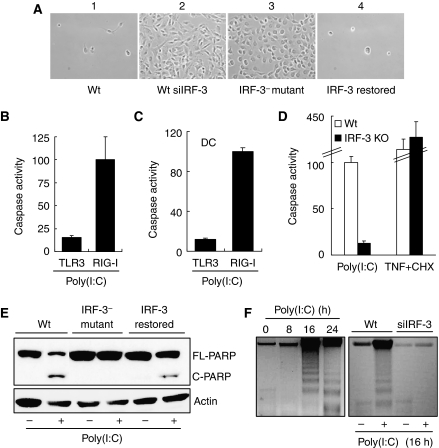
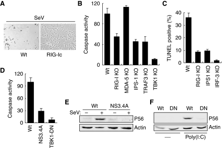
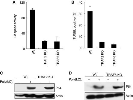
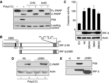
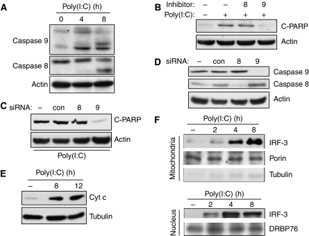
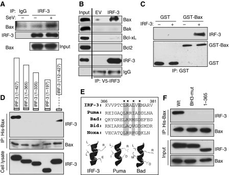
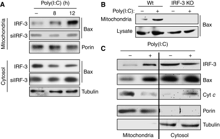
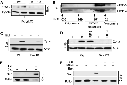
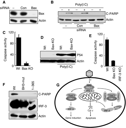
Upon infection with many RNA viruses, the cytoplasmic retinoic acid inducible gene-I (RIG-I) pathway activates the latent transcription factor IRF-3, causing its nuclear translocation and the induction of many antiviral genes, including those encoding interferons. Here, we report a novel and distinct activity of IRF-3, in virus-infected cells, that induces apoptosis. Using genetically defective mouse and human cell lines, we demonstrated that, although both pathways required the presence of RIG-I, IPS1, TRAF3 and TBK1, only the apoptotic pathway required the presence of TRAF2 and TRAF6 in addition. More importantly, transcriptionally inactive IRF-3 mutants, such as the one missing its DNA-binding domain, could efficiently mediate apoptosis. Apoptosis was triggered by the direct interaction of IRF-3, through a newly identified BH3 domain, with the pro-apoptotic protein Bax, their co-translocation to the mitochondria and the resulting activation of the mitochondrial apoptotic pathway. Thus, IRF-3 is a dual-action cytoplasmic protein that, upon activation, translocates to the nucleus or to the mitochondrion and triggers two complementary antiviral responses of the infected cell.


Conflict of interest statement

The authors declare that they have no conflict of interest.

Figures

Figure 1
Figure 1
IRF-3 activation by cytoplasmic dsRNA signalling triggers apoptosis. (A) HT1080-derived cell lines were infected with Sendai virus and 3 days after infection culture fields were photographed. 1, U4C cells (Wt); 2, IRF-3-knocked-down HT1080 cells (Wt-siIRF-3); 3, P2.1 cells (IRF-3 mutant); 4, IRF-3-restored P2.1 cells. (B) HT1080 cells were treated (TLR3) or transfected (RIG-I) with poly(I:C) for 16 h when caspase activation was measured. (C) Primary bone-marrow-derived dendritic cells (DCs), isolated from Wt C57Bl/6 mice, were treated (TLR3) or transfected (RIG-I) with poly(I:C); caspase activity was measured 16 h after treatment. (D) Wt and IRF-3 KO MEFs were transfected with 8 μg/ml of poly(I:C) or treated with 100 ng/ml of TNF and 1 μg/ml of cycloheximide (CHX) for 16 h, when caspase activation was measured; fold induction of caspase activity of Wt MEFs was considered as 100 and all other values were normalized to this. (E) Wt, IRF-3 mutant and IRF-3-restored cells (as described in (A)) were transfected with poly(I:C) and PARP cleavage was analysed 16 h after transfection by western blot. (F) Wt and Wt-siIRF-3 cells (as described in (A)) were transfected with poly(I:C) for the indicated time, when cellular DNA was isolated and analysed by running on 1.5% agarose gel. The left panel shows kinetics of DNA fragmentation for Wt cells. For the above experiments, 2 μg/ml of poly(I:C) was transfected (B, C, E, F) or 100 μg/ml of poly(I:C) was added into the culture medium in (B, C); fold induction of caspase activity by RIG-I signalling in response to transfected poly(I:C) was considered as 100 and other values were normalized to this (B, C).
Figure 2
Figure 2
Components of RIG-I signalling are required for apoptosis. (A) HT1080 cells (Wt) and HT1080 cells expressing a dominant-negative mutant of RIG-I (RIG-Ic) were infected with SeV; 3 days after infection culture fields were photographed. (B) Wt and KO MEF cells (as indicated) were transfected with poly(I:C), caspase activity was measured after 16 h; fold induction of caspase activity of Wt MEFs was considered as 100 and all other values were normalized to this. (C) Wt and knockout MEF cells (as indicated), grown on coverslips, were transfected with poly(I:C) for 16 h, when the cells were stained with FITC-conjugated TUNEL; TUNEL-positive and DAPI-stained cells were counted under the microscope and percent TUNEL-positive cells are presented. (D) Human cells expressing Hepatitis C virus protease NS3.4A or HT1080 cells expressing a dominant-negative mutant of TBK1 (TBK1-DN) were used for this experiment. The cells were transfected with poly(I:C), caspase activity was measured 16 h after transfection. (E) Cell lysates from NS3.4A-expressing cells (as described in (C)) were analysed for induction of P56 by SeV infection (6 h after infection) by western blot. (F) TBK1-DN (DN)-expressing cells (as described in (C)) were analysed for induction of P56 by western blot, 6 h after poly(I:C) transfection.
Figure 3
Figure 3
Additional components are required for apoptotic pathway. (A) Wt, TRAF2 KO and TRAF6 KO MEFs were transfected with poly(I:C); caspase activity was measured 16 h after transfection. (B) Wt and KO MEF cells (as indicated) were transfected with poly(I:C); 16 h after transfection the cells were stained with FITC-conjugated TUNEL; TUNEL-positive and DAPI-stained cells were counted under the microscope and the percentage of positive cells are shown here. (C, D) Wt and KO MEF cells (as indicated) were transfected with poly(I:C); induction of P54 was analysed 12 h after transfection, by western blot.
Figure 4
Figure 4
dsRNA-signalling-mediated apoptosis is independent of IRF-3-driven gene induction. (A) HT1080 cells were transfected with poly(I:C) in the presence or absence of the protein synthesis inhibitor, cycloheximide, or the RNA synthesis inhibitor, actinomycin D, for 12 h, when the cell extracts were analysed for cleaved PARP and P56 induction by western blot. (B) A schematic representation of IRF-3; DNA-binding domain (DBD), nuclear localization signal (NLS), nuclear export signal (NES) and critical phosphoacceptor sites on the C-terminus are indicated. (C) HT1080-siIRF-3 cells expressing Wt IRF-3 (Wt), IRF-3 396/98AA mutant (396AA), IRF-3 385/86AA mutant (385AA) or IRF-3 386A (a stable pool of cells) were transfected with poly(I:C); caspase activity was measured after 16 h and the cell lysates were analysed for the expression of IRF-3 mutant proteins. (D, E) HT1080-siIRF-3 cells expressing Wt or DBD-deleted IRF-3 mutant (ΔDBD, IRF-3 residues 1–112 were deleted, as in (A)) were transfected with poly(I:C) and the cell extracts were analysed for cleaved PARP (16 h after transfection) or P56 induction (6 h after transfection) by western blot.
Figure 5
Figure 5
dsRNA-induced apoptosis is accompanied by mitochondrial translocation of IRF-3. (A) HT1080 cells were transfected with poly(I:C) for the indicated time, when the cell extracts were analysed for the activation of caspase 8 and caspase 9 by western blot. (B) HT1080 cells were transfected with poly(I:C) in the presence or absence of caspase inhibitors (caspase 8 inhibitor, z-IETD-FMK, and caspase 9 inhibitor, z-LEHD-FMK, as indicated); cell extracts were analysed for cleaved PARP by western blot. (C, D) HT1080 cells were transfected with siRNAs specific for caspase 8, caspase 9 or a control siRNA against cyclophilin A (Dharmacon); 48 h after transfection, the cells were transfected with poly(I:C) and analysed for PARP cleavage or, as indicated, by western blot. (E) Wt IRF-3-expressing cells were transfected with poly(I:C) for the indicated time, when the cytosolic fractions were analysed for Cyt c release by western blot. (F) Wt IRF-3-expressing cells (doubly tagged with N-terminal V5 and C-terminal Flag) were transfected with poly(I:C) for the indicated time, when the mitochondrial and nuclear fractions were isolated and analysed for the presence of IRF-3 and other proteins, as indicated, by western blot.
Figure 6
Figure 6
IRF-3 interacts with Bax using a BH3-like domain on its C-terminus. (A) HT1080-siIRF-3 cells expressing Wt IRF-3 were infected with SeV; 2 h after infection IRF-3 was immunoprecipitated and analysed for Bax by western blot. (B) HT1080-siIRF-3 cells expressing Wt IRF-3 (or empty vector, EV) were transfected with poly(I:C) for 2 h and the cell lysates were immunoprecipitated with IRF-3 antibody and the immunoprecipitates were analysed for the indicated proteins by western blot. (C) IRF-3 was purified from human cells as described in Materials and methods and used for in vitro interaction assay with recombinant GST-conjugated human Bax (ABNOVA) and the reaction mixture was pulled down with GST beads and analysed for IRF-3 by western blot. (D) HT1080-siIRF-3 cells were transiently co-transfected with N-terminally V5-tagged Wt or deletion mutants of IRF-3 (as indicated) and N-terminally His-tagged Bax; 16 h after transfection cells were transfected with dsRNA for 2 h, when the cell lysates were treated with Ni-NTA and bound proteins were analysed for IRF-3 by western blot. (E) Human IRF-3 residues (366–381) were aligned with the BH3 domains of pro-apoptotic proteins (as indicated) and a molecular modelling of this domain is shown in comparison with the BH3 domains of Puma and Bad. (F) HT1080-siIRF-3 cells were co-transfected with Wt or mutants of IRF-3 (BH-3 domain mutant (BH3-mut)), wherein the critical residues (as shown by * in (E)) were mutated to Ala, (1–365) mutant, IRF-3 mutant consisting of residues 1–365, and human Bax (as in (D)); the cells were transfected with poly(I:C) for 2 h, when the cell lysates were treated with Ni-NTA and bound proteins were analysed for IRF-3 by western blot.
Figure 7
Figure 7
IRF-3 interaction translocates Bax to the mitochondria. (A) HT1080-siIRF-3 (siIRF-3) or IRF-3-restored cells (IRF-3) were transfected with poly(I:C); the mitochondrial and the cytosolic fractions were analysed by western blot for the indicated proteins. (B) Wt and IRF-3 KO MEFs were transfected with poly(I:C); the mitochondrial fractions and the cell lysates were analysed by western blot. (C) HT1080-siRIF-3 cells expressing IRF-3 were transfected with poly(I:C); the mitochondrial and cytosolic fractions were isolated and analysed as indicated.
Figure 8
Figure 8
IRF-3 interaction activates Bax and causes Cyt c release from the mitochondria. (A) HT1080 (Wt) and HT1080-siIRF-3 (siIRF-3) cells were transfected with poly(I:C) for 6 h, after which the cell lysates were immunoprecipitated with Bax 6A-7 antibody and the immunoprecipitates were analysed by western blot using polyclonal Bax antibody. (B) Purified human IRF-3 was used for Bax oligomerization assay as described in Materials and methods; at the end of the reaction, the mitochondrial pellet was extracted in HE buffer containing 1% CHAPS and the extract was applied to Superdex 200 PG XK16/60 column; the fractions were analysed by western blot for Bax; column calibration was performed using the molecular weight standards kit as indicated. (C) Caspase-8-cleaved Bid (Bid, R&D Systems, 50 nM) was used for mitochondrial activation assay using the mitochondria-free cytosolic extract from Wt or Bax KO MEF cells as described in Materials and methods; at the end of the reaction, the mitochondrial pellet and the supernatant (sup) were analysed for released Cyt c, as indicated. (D) Purified activated human IRF-3 (500 nM) was used in mitochondrial activation assay using the mitochondria-free cytosolic extract from Wt or Bax KO MEF cells and analysed as described in (C). (E) Bid (50 nM) was used for in vitro Cyt c release assay using recombinant human Bax (120 nM) as described in Materials and methods; at the end of the reaction, the supernatant (sup) and the pellet were analysed for Cyt c by western blot. (F) Purified activated IRF-3 (500 nM) was used for in vitro Cyt c release assay using recombinant human Bax (120 nM) and analysed as described in (E).
Figure 9
Figure 9
Bax is essential for dsRNA-induced apoptosis. (A) HT1080 cells were transfected with 100 nmol/ml of Bax siRNA or a control siRNA (against cyclophilin A) (Dharmacon) using DharmaFECT 4 reagent; cell extracts were subjected to western blot analysis after 48 h. (B) RNAi-mediated knockdown of Bax was carried out by transfection of HT1080 cells as in (A); 48 h later, these cells were transfected with poly(I:C) and the cell extracts were analysed for cleaved PARP by western blot. (C) Wt and Bax KO MEFs were transfected with poly(I:C); caspase activation was measured after 16 h. (D) Wt and Bax KO MEF cells were transfected with poly(I:C) and cell extracts were analysed for P54 induction by western blot. (E) Primary bone-marrow-derived DCs were isolated from Wt, IRF-3 KO and Bax KO mice and infected with SeV (at MOI: 10) for 72 h, when the cell lysates were analysed for caspase activity. (F) HT1080-siIRF-3 cells expressing Wt or mutants of IRF-3 (as in Figure 6D; a stable pool was generated) were transfected with poly(I:C) for 16 h, when the cell lysates were analysed for PARP cleavage. (G) A model showing the transcriptional and apoptotic pathways mediated by dual actions of IRF-3.

Comment in

References

    1. Alexopoulou L, Holt AC, Medzhitov R, Flavell RA (2001) Recognition of double-stranded RNA and activation of NF-kappaB by Toll-like receptor 3. Nature 413: 732–738 - PubMed
    1. Barber GN (2001) Host defense, viruses and apoptosis. Cell Death Differ 8: 113–126 - PubMed
    1. Der SD, Yang YL, Weissmann C, Williams BR (1997) A double-stranded RNA-activated protein kinase-dependent pathway mediating stress-induced apoptosis. Proc Natl Acad Sci USA 94: 3279–3283 - PMC - PubMed
    1. Doyle S, Vaidya S, O'Connell R, Dadgostar H, Dempsey P, Wu T, Rao G, Sun R, Haberland M, Modlin R, Cheng G (2002) IRF3 mediates a TLR3/TLR4-specific antiviral gene program. Immunity 17: 251–263 - PubMed
    1. Elco CP, Guenther JM, Williams BR, Sen GC (2005) Analysis of genes induced by Sendai virus infection of mutant cell lines reveals essential roles of interferon regulatory factor 3, NF-kappaB, and interferon but not toll-like receptor 3. J Virol 79: 3920–3929 - PMC - PubMed

Publication types