. 2010 May 5;29(9):1585-99.
doi: 10.1038/emboj.2010.43. Epub 2010 Apr 1.

AIF promotes chromatinolysis and caspase-independent programmed necrosis by interacting with histone H2AX

Affiliations

AIF promotes chromatinolysis and caspase-independent programmed necrosis by interacting with histone H2AX

Cédric Artus et al. EMBO J. .

Abstract

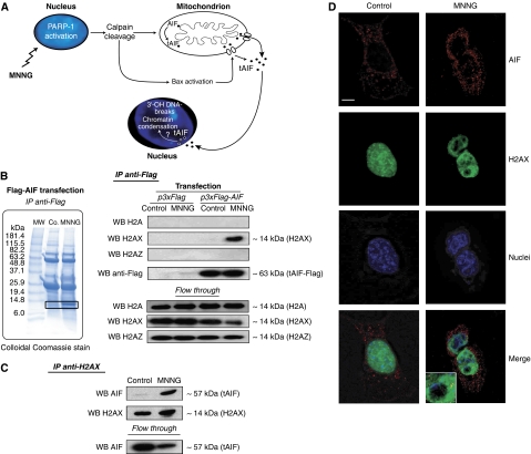
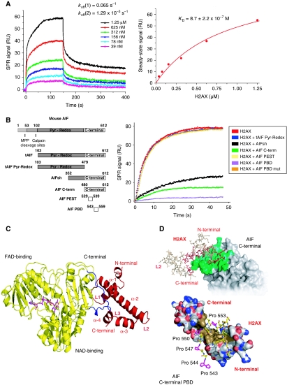
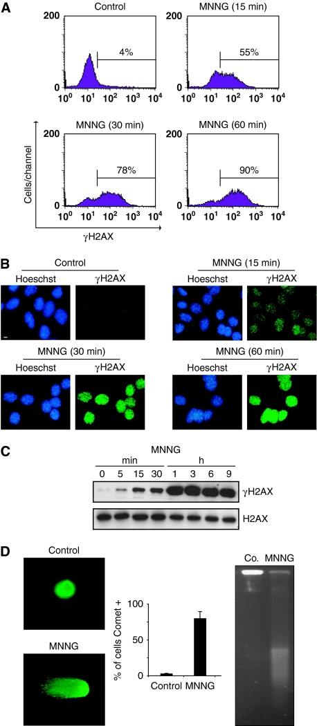
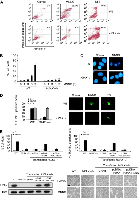
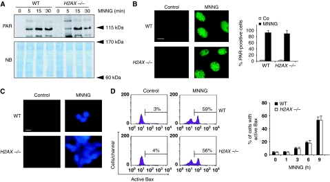
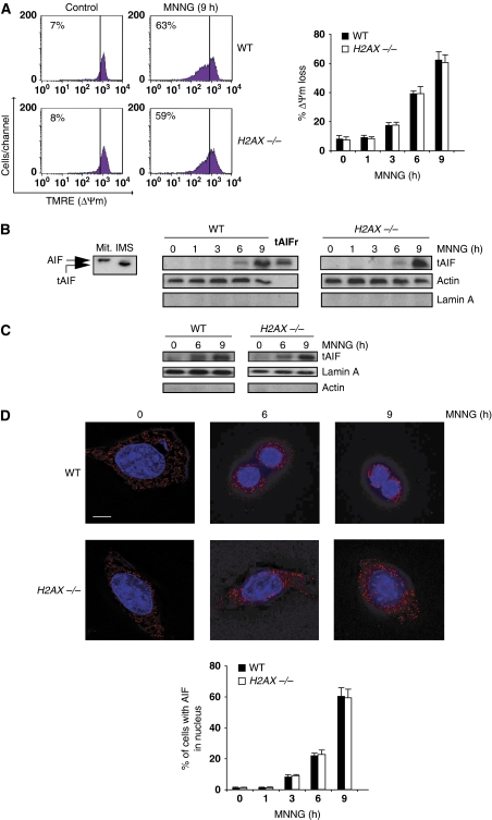
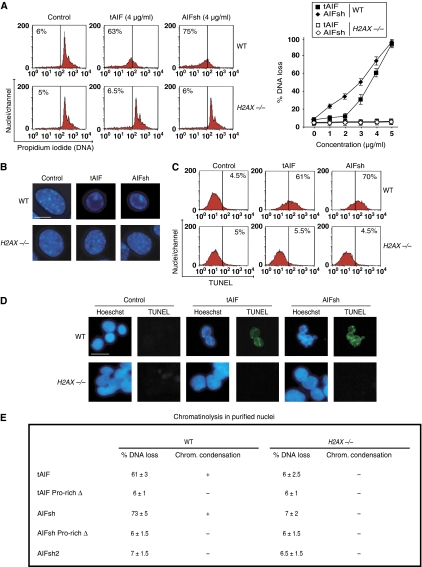
Programmed necrosis induced by DNA alkylating agents, such as MNNG, is a caspase-independent mode of cell death mediated by apoptosis-inducing factor (AIF). After poly(ADP-ribose) polymerase 1, calpain, and Bax activation, AIF moves from the mitochondria to the nucleus where it induces chromatinolysis and cell death. The mechanisms underlying the nuclear action of AIF are, however, largely unknown. We show here that, through its C-terminal proline-rich binding domain (PBD, residues 543-559), AIF associates in the nucleus with histone H2AX. This interaction regulates chromatinolysis and programmed necrosis by generating an active DNA-degrading complex with cyclophilin A (CypA). Deletion or directed mutagenesis in the AIF C-terminal PBD abolishes AIF/H2AX interaction and AIF-mediated chromatinolysis. H2AX genetic ablation or CypA downregulation confers resistance to programmed necrosis. AIF fails to induce chromatinolysis in H2AX or CypA-deficient nuclei. We also establish that H2AX is phosphorylated at Ser139 after MNNG treatment and that this phosphorylation is critical for caspase-independent programmed necrosis. Overall, our data shed new light in the mechanisms regulating programmed necrosis, elucidate a key nuclear partner of AIF, and uncover an AIF apoptogenic motif.


Conflict of interest statement

The authors declare that they have no conflict of interest.

Figures

Figure 1
Figure 1
AIF interacts with histone H2AX in MNNG-induced programmed necrosis. (A) Model for alkylating DNA damage-mediated death. MNNG induces death through PARP-1, calpains, and Bax activation. These proteins facilitate the cleavage and release of tAIF (the pro-apoptotic AIF form) from the mitochondrion to the cytosol and nucleus where tAIF generates 3′-OH DNA breaks and chromatin condensation through a yet unknown mechanism. (B) Representative SDS–PAGE and Coomassie blue staining of Flag-immunoprecipitates (IP anti-Flag) obtained from MEFs transfected with Flag-AIF and either untreated (Co.) or treated with MNNG (9 h). MW, molecular weight marker. After MNNG treatment, a 14 kDa protein (in the square) was co-purified with AIF. Flag-immunoprecipitates (IP anti-Flag) obtained from MEFs transfected with Flag-AIF and either untreated (Co.) or treated with MNNG (9 h) were analysed by western blotting (WB) using anti-H2A, anti-H2AX, anti-H2AZ, and anti-Flag antibodies. WB on lysates after IP is also shown (flow through). (C) H2AX immunoprecipitation on MEFs incubated or not with MNNG (9 h). AIF and H2AX were detected by immunoblot. WB on lysates after IP is also shown (flow through). (D) MEFs left untreated (control) or treated with MNNG as above were subjected to confocal immunofluorescent detection of AIF (red) and H2AX (green). Nuclei were stained with Hoechst 33342 (blue). This experiment was repeated five times, yielding similar results. Bar: 10 μm.
Figure 2
Figure 2
AIF interacts with H2AX through its C-terminal PBD. (A) SPR sensorgrams resulting from the injection of the indicated concentrations of soluble H2AX onto immobilized tAIF. koff, dissociation rate constants. Right: H2AX concentration dependence of the steady-state response. KD, equilibrium dissociation constant. (B) Mouse AIF, tAIF, tAIF Pyr-Redox, AIFsh, AIF C-term, AIF PEST, and AIF PBD protein organization (Left). Numbers designate aminoacids. Pyr-Redox and C-terminal AIF domains are indicated. Right: SPR sensorgrams showing the association of H2AX with immobilized tAIF, in the absence or presence of tAIF Pyr-Redox, AIFsh, AIF C-term, AIF PEST, AIF PBD, or AIF PBD mut. (C) Model of the tAIF/H2AX complex obtained by homology with the known crystal structures of the individual partners. Left: ribbon structure of tAIF (yellow) and H2AX (red). AIF interacts with H2AX through its C-terminal residues 543–559 (in blue). FAD (the AIF cofactor) is drawn in magenta. H2AX folding consists of four α helices (α-1 to α-4) linked by three short loops (L1–L3). (D) tAIF/H2AX molecular docking. Upper: surface of the tAIF binding interface (green). Residues of H2AX interacting with tAIF surface are drawn in red. Lower: tAIF C-terminal proline-rich residues interact with H2AX through a hydrophobic cluster (brown). Acidic and basic residues are, respectively, depicted in red and blue. AIF C-terminal Pro543, 544, 547, 550, and 553 are drawn in magenta.
Figure 3
Figure 3
MNNG induces H2AX phosphorylation and DNA double-strand breaks. (A) Kinetics of the H2AX phosphorylation induced by MNNG in MEFs. Cells were left untreated (control) or treated with MNNG, and H2AX Ser139 phosphorylation was assessed by flow cytometry. Representative cytofluorometric plots are shown. Percentages refer to γH2AX-positive staining. This experiment was repeated five times with a lower experimental variability (⩽5%). (B) Immunofluorescent staining of γH2AX detected in MEFs left untreated (control) or treated with MNNG and stained with Hoechst 33342 (to visualize nuclei) or a specific γH2AX antibody. Representative cells are shown. This experiment was repeated eight times with similar results. Bar: 10 μm. (C) γH2AX immunoblotting detection in total extracts obtained at different times from MEFs untreated or treated with MNNG. The membrane was reblotted for total H2AX to control protein loading. (D) Analysis of DNA DSB by a neutral comet assay performed in MEFs untreated or treated with MNNG (1 h). Representative comet images and percentage of cells with a tail (comet +) are shown. Data are means±s.d. (n=6). Alternatively, MEFs were analysed by field inversion gel electrophoresis (FIGE). The smear observed in MNNG-treated cells indicated accumulation of DSB (Saintigny et al, 2001). A full-colour version of this figure is available at The EMBO Journal Online.
Figure 4
Figure 4
γH2AX is essential for programmed necrosis. (A) WT and H2AX−/− cells were untreated (control) or treated with MNNG (9 h) or staurosporine (STS), labelled with AnnexinV-FITC and PI, and analysed by flow cytometry. Representative cytofluorometric plots are shown. Percentages refer to double-positive staining. (B) Kinetic analysis of PS exposure and cell viability loss induced by MNNG in WT and H2AX−/− MEFs. After the indicated times, cells were stained as in (A) and the frequency of double-positive labelling was recorded and expressed as a percentage. Data are the means of 10 independent experiments±s.d. (C) WT and H2AX−/− MEFs were treated with MNNG (9 h) and stained with Hoechst 33342 to visualize nuclei. Representative nuclei from untreated (control) or MNNG-treated cells are shown. Bar: 10 μm. (D) WT and H2AX−/− MEFs were untreated (Co) or treated with MNNG (9 h) or STS as in (A), stained for the detection of 3′-OH DNA breaks, and analysed by flow cytometry. Data are the means of six independent experiments±s.d. Right: representative microphotographs from each treatment are shown. Bar: 10 μm. (E) H2AX−/− MEFs were transfected with the indicated expression plasmids and selected as described in ‘Materials and methods' section. Then, cells were untreated (Co) or treated with MNNG (9 h). Cell death was analysed by AnnexinV-FITC/PI and TUNEL staining. Data are means±s.d. (n=8). The expression level of H2AX was assessed by immunoblotting. Equal loading was confirmed by histone H2A assessment. Representative microphotographs of each treatment are shown. Bar: 40 μm. A full-colour version of this figure is available at The EMBO Journal Online.
Figure 5
Figure 5
PARP-1, calpains, and Bax are activated in the absence of H2AX. (A) Poly(ADP-ribose) (PAR) immunoblotting detection in total extracts from WT and H2AX−/− MEFs untreated or treated with MNNG at different times. The membrane was stained with naphtol blue (NB) to assess protein loading. (B) WT and H2AX−/− MEFs were untreated (control) or treated with MNNG (15 min), immunostained for PAR detection, and visualized by fluorescent microscopy. Representative micrographs of each cell type are shown. Data are means±s.d. (n=5). Bar: 10 μm. (C) Fluorescent assessment of calpain activity measured in WT and H2AX−/− MEFs untreated (control) or treated with MNNG (1 h). Representative micrographs of each treatment are shown. This experiment was repeated four times, yielding similar results. Bar: 10 μm. (D) WT and H2AX−/− MEFs were treated with MNNG and Bax activation was measured by flow cytometry. Representative cytofluorometric plots of untreated (control) and MNNG-treated (9 h) cells are shown. Data are the means±s.d. (n=6). A full-colour version of this figure is available at The EMBO Journal Online.
Figure 6
Figure 6
MNNG-induced ΔΨm loss and tAIF relocalization from the mitochondria to the cytosol and nucleus in H2AX−/− MEFs. (A) After the indicated time post-MNNG treatment, WT and H2AX−/− MEFs were labelled with TMRE, and the frequency of cells with ΔΨm loss was recorded and illustrated as a plot. Data presented in the bar chart are from the means of five independent experiments±s.d. In cytometry panels, percentages refer to cells with ΔΨm loss. (B) Mitochondrial AIF (Mit.; 62 kDa) is cleaved into a lower molecular weight tAIF (57 kDa) in the mitochondrial intermembrane space (IMS) upon atractyloside treatment (Yuste et al, 2005) (left panel). Cytosolic fractions recovered from WT and H2AX−/− MEFs after MNNG treatment at different times were blotted for tAIF detection. Compared to the mitochondria isolated from WT MEFs and tAIF recombinant protein (tAIFr), MNNG treatment induces both AIF cleavage into tAIF and time-dependent tAIF release to cytosol. Lamin A and actin were used to control fractionation quality and protein loading. (C) tAIF was also detected in nuclear extracts obtained from WT and H2AX−/− MEFs treated as in (B). Actin and lamin A were used to control fractionation quality and protein loading. (D) WT and H2AX−/− MEFs were untreated or treated with MNNG (6 and 9 h), immunostained for AIF detection (red), and examined by confocal microscopy. Hoechst 33342 (blue) was used to visualize nuclei. Representative microphotographs are shown. Bar: 10 μm. Cells presenting AIF in the nucleus are quantified and plotted as a percentage of total cells. This experiment was repeated four times, yielding similar results.
Figure 7
Figure 7
Cooperative effect of AIF and H2AX on chromatinolysis. (A) Purified WT and H2AX−/− MEFs nuclei were incubated in the absence (control) or presence of mouse recombinant proteins tAIF and AIFsh and stained with PI. The percentage of DNA loss was measured by flow cytometry and illustrated as a plot. Data are shown as mean values±s.d. (n=4). In cytometry panels, numbers indicate the % of hypoploid nuclei. (B) WT and H2AX−/− MEFs nuclei were treated with tAIF and AIFsh (4 μg/ml) and stained with Hoescht 33342 to visualize chromatin condensation by fluorescent microscopy. Representative micrographs of each treatment are shown. This experiment was done three times, yielding similar results. (C) Nuclei were treated with tAIF and AIFsh as in (B), stained for the detection of 3′-OH DNA breaks, and analysed by flow cytometry. Representative cytofluorometric plots are shown. (D) In a similar experiment to (C), nuclei were stained with Hoechst 33342, and blue and green (TUNEL positive) fluorescence was visualized. Representative microphotographs from untreated (control), tAIF, or AIFsh-treated nuclei are shown. Bar: 20 μm. (E) Table compiling the results obtained in WT and H2AX−/− MEFs nuclei incubated in the presence of tAIF, tAIF Pro-rich Δ, AIFsh, AIFsh Pro-rich Δ, and AIFsh2 (4 μg/ml). Data are represented as % of DNA loss measured as in (A) (X±s.d.; n=5), +: induction of chromatin condensation, or −: absence of chromatin condensation. A full-colour version of this figure is available at The EMBO Journal Online.
Figure 8
Figure 8
The AIF/H2AX link promotes DNA degradation by activating CypA. (A) WT MEFs were transfected with a scramble siRNA (control siRNA) or two different siRNAs against mouse CypA (CypAsiRNAa and CypAsiRNAb) or mouse EndoG (EndoGsiRNAa and EndoGsiRNAb). Total cell lysates from siRNA cells were prepared and the expression levels of CypA or EndoG were assessed by immunoblotting. Actin was used as a loading control; 48 h after the indicated transfection, cells were untreated (control) or treated with MNNG (9 h), labelled with AnnexinV-FITC and PI, and analysed by flow cytometry. Representative cytofluorometric plots are shown. Percentages refer to double-positive staining. Kinetic analysis of PS exposure and cell viability loss induced by MNNG. After the indicated times, cells were stained as above and the frequency of double-positive labelling was recorded and expressed as a percentage. Data are the means of eight independent experiments±s.d. (B) MEFs were untreated or treated with MNNG as in (A), stained for the detection of 3′-OH DNA breaks, and analysed by flow cytometry. Data are the means of six independent experiments±s.d. (C) MEFs were untreated or treated with MNNG (9 h) and stained with Hoechst 33342 to visualize nuclei. The number of cells presenting chromatin condensation were quantified and plotted as a percentage of total cells. Data are the means±s.d. (n=6). The asterisks in (AC) indicate a significant effect (P<0.01). (D) Nuclei purified from the MEFs described in (A) were incubated in the absence (control) or presence of mouse recombinant proteins tAIF or tAIF Pro-rich Δ and stained with PI. The percentage of DNA loss was measured by flow cytometry, numbers indicate the % of hypoploid nuclei. In selected experiments, nuclei purified from WT CypAsiRNAa MEFs were reconstituted with recombinant CypA (10 nM). A full-colour version of this figure is available at The EMBO Journal Online.

References

    1. Artus C, Maquarre E, Moubarak RS, Delettre C, Jasmin C, Susin SA, Robert-Lezenes J (2006) CD44 ligation induces caspase-independent cell death via a novel calpain/AIF pathway in human erythroleukemia cells. Oncogene 25: 5741–5751 - PubMed
    1. Banath JP, Olive PL (2003) Expression of phosphorylated histone H2AX as a surrogate of cell killing by drugs that create DNA double-strand breaks. Cancer Res 63: 4347–4350 - PubMed
    1. Bewersdorf J, Bennett BT, Knight KL (2006) H2AX chromatin structures and their response to DNA damage revealed by 4Pi microscopy. Proc Natl Acad Sci USA 103: 18137–18142 - PMC - PubMed
    1. Bonner WM, Redon CE, Dickey JS, Nakamura AJ, Sedelnikova OA, Solier S, Pommier Y (2008) GammaH2AX and cancer. Nat Rev Cancer 8: 957–967 - PMC - PubMed
    1. Cande C, Vahsen N, Kouranti I, Schmitt E, Daugas E, Spahr C, Luban J, Kroemer RT, Giordanetto F, Garrido C, Penninger JM, Kroemer G (2004) AIF and cyclophilin A cooperate in apoptosis-associated chromatinolysis. Oncogene 23: 1514–1521 - PubMed

Publication types

MeSH terms