. 2010 Apr 1;464(7289):763-7.
doi: 10.1038/nature08855.

Impaired hippocampal-prefrontal synchrony in a genetic mouse model of schizophrenia

Affiliations

Impaired hippocampal-prefrontal synchrony in a genetic mouse model of schizophrenia

Torfi Sigurdsson et al. Nature. .

Abstract

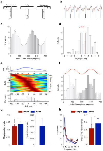
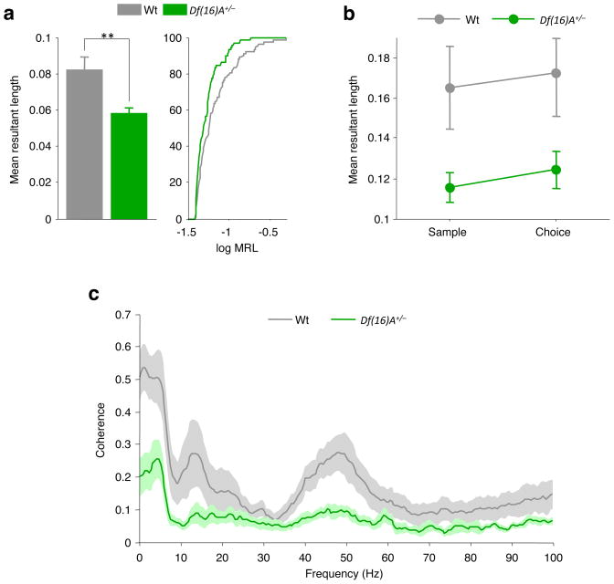
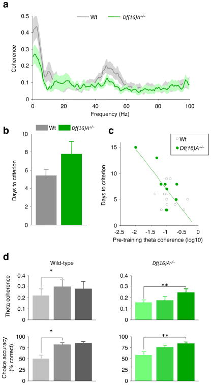
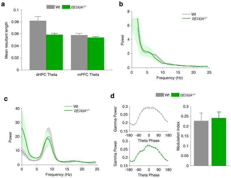
Abnormalities in functional connectivity between brain areas have been postulated as an important pathophysiological mechanism underlying schizophrenia. In particular, macroscopic measurements of brain activity in patients suggest that functional connectivity between the frontal and temporal lobes may be altered. However, it remains unclear whether such dysconnectivity relates to the aetiology of the illness, and how it is manifested in the activity of neural circuits. Because schizophrenia has a strong genetic component, animal models of genetic risk factors are likely to aid our understanding of the pathogenesis and pathophysiology of the disease. Here we study Df(16)A(+/-) mice, which model a microdeletion on human chromosome 22 (22q11.2) that constitutes one of the largest known genetic risk factors for schizophrenia. To examine functional connectivity in these mice, we measured the synchronization of neural activity between the hippocampus and the prefrontal cortex during the performance of a task requiring working memory, which is one of the cognitive functions disrupted in the disease. In wild-type mice, hippocampal-prefrontal synchrony increased during working memory performance, consistent with previous reports in rats. Df(16)A(+/-) mice, which are impaired in the acquisition of the task, showed drastically reduced synchrony, measured both by phase-locking of prefrontal cells to hippocampal theta oscillations and by coherence of prefrontal and hippocampal local field potentials. Furthermore, the magnitude of hippocampal-prefrontal coherence at the onset of training could be used to predict the time it took the Df(16)A(+/-) mice to learn the task and increased more slowly during task acquisition. These data suggest how the deficits in functional connectivity observed in patients with schizophrenia may be realized at the single-neuron level. Our findings further suggest that impaired long-range synchrony of neural activity is one consequence of the 22q11.2 deletion and may be a fundamental component of the pathophysiology underlying schizophrenia.


Figures

Figure 1
Figure 1. Hippocampal–prefrontal synchrony during a spatial working memory task in mice
a, Spatial working memory task (see text). R, reward. b, Example field potential recording from the hippocampus (black trace) showing theta oscillations (red trace) and spikes recorded simultaneously from a prefrontal neuron (tick marks). Scale bar, 200 ms. c, Distribution of hippocampal theta phases at which the neuron in b fired. d, Distribution of Rayleigh z scores, for estimating significance of phase-locking to hippocampal theta oscillations, for prefrontal neurons recorded from wild-type mice. The red line denotes the significance threshold (P < 0.05); 83 of 125 neurons were significantly phase-locked. e, Prefrontal neurons phase-lock most strongly to the theta phase of the past. Top: strength of phase-locking of a single prefrontal neuron as a function of lag between the prefrontal spike and the hippocampal field potential recording. MRL, mean resultant length (Methods). Middle: normalized phase-locking strength of each neuron as a function of lag. Rows represent individual neurons ordered by the lag of maximal phase-locking. Arrow, neuron corresponding to trace at top. Bottom: distribution of lags at which each cell was maximally phase-locked (lag, −17.5 ± 6.2 ms (mean ± s.e.m.); P = 0.016; Wilcoxon signed-rank test). f, Distribution of hippocampal theta phases to which prefrontal neurons are significantly phase-locked (mean ± 95% confidence interval, 166° ± 43.8°; P = 0.0005, Rayleigh test). The red trace is a schematic of a single theta cycle. g, Phase-locking strength in the centre arm of the maze during sample and choice phases (left) and the difference in phase-locking across the two conditions (right). h, Coherence between hippocampal and prefrontal field potentials during the same task phases as in g: an example of coherence in one session (left); theta coherence (4–12 Hz) across animals (right). *P < 0.05; data shown, mean ± s.e.m.
Figure 2
Figure 2. Reduced hippocampal–prefrontal synchrony in Df(16)A+/− mice
a, Phase-locking of prefrontal neurons to hippocampal theta (left) and cumulative distribution of phase-locking values (right) in the two genotypes. Phase-locking is stronger in wild-type mice. **P < 0.01; WT, wild type. b, Phase-locking strength in the centre arm during sample and choice phases. c, Coherence between hippocampal and prefrontal field potentials during the same task phases as in b. Coherence is lower in Df(16)A+/− mice. Data shown, mean ± s.e.m.
Figure 3
Figure 3. Reduced hippocampal–prefrontal synchrony correlates with behavioural performance in Df(16)A+/− mice
a, Coherence between the prefrontal cortex and the hippocampus during habituation sessions before training on the spatial working memory task. b, Days taken to reach criterion performance on the spatial working memory task. c, Days taken to reach criterion versus theta coherence during habituation sessions for each animal. Animals with lower theta coherence before training take longer to learn the spatial working memory task. Green line, linear regression of data from Df(16)A+/− mice d, Development of hippocampal–prefrontal coherence during acquisition of the working memory task: theta coherence (top) and choice accuracy (bottom) during early (trials 1–5), middle (trials 26–30) and late (session in which criterion was reached) stages of training in wild-type (left) and Df(16)A+/− (right) mice. *P < 0.05, **P < 0.01. Data shown, mean ± s.e.m.
Figure 4
Figure 4. Selectivity of hippocampal–prefrontal synchrony deficits in Df(16)A+/− mice
a, Comparison of phase-locking to theta oscillations in prefrontal cortex and hippocampus across the two genotypes. Phase-locking to prefrontal theta oscillations is intact. b, c, Field potential power in the prefrontal cortex (b) and hippocampus (c). d, Modulation of hippocampal gamma (30–80-Hz) power by theta phase. Left: examples of theta–gamma modulation in a wild-type mouse (top) and in a Df(16)A+/− mouse (bottom). Right: mean theta–gamma modulation index (Methods). Data shown, mean ± s.e.m.

References

    1. Stephan KE, Friston KJ, Frith CD. Dysconnection in schizophrenia: from abnormal synaptic plasticity to failures of self-monitoring. Schizophr Bull. 2009;35:509–527. - PMC - PubMed
    1. Wernicke C. Grundrisse der Psychiatrie. Thieme; 1906.
    1. Meyer-Lindenberg AS, et al. Regionally specific disturbance of dorsolateral prefrontal-hippocampal functional connectivity in schizophrenia. Arch Gen Psychiatry. 2005;62:379–386. - PubMed
    1. Lawrie SM, et al. Reduced frontotemporal functional connectivity in schizophrenia associated with auditory hallucinations. Biol Psychiatry. 2002;51:1008–1011. - PubMed
    1. Gottesman II, Shields J. A polygenic theory of schizophrenia. Proc Natl Acad Sci USA. 1967;58:199–205. - PMC - PubMed

Publication types

MeSH terms