. 2010 Mar 25;5(3):e9870.
doi: 10.1371/journal.pone.0009870.

Tgf-Beta inhibition restores terminal osteoblast differentiation to suppress myeloma growth

Affiliations

Tgf-Beta inhibition restores terminal osteoblast differentiation to suppress myeloma growth

Kyoko Takeuchi et al. PLoS One. .

Abstract

Background: Multiple myeloma (MM) expands almost exclusively in the bone marrow and generates devastating bone lesions, in which bone formation is impaired and osteoclastic bone resorption is enhanced. TGF-beta, a potent inhibitor of terminal osteoblast (OB) differentiation, is abundantly deposited in the bone matrix, and released and activated by the enhanced bone resorption in MM. The present study was therefore undertaken to clarify the role of TGF-beta and its inhibition in bone formation and tumor growth in MM.

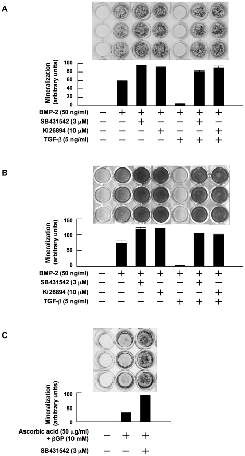
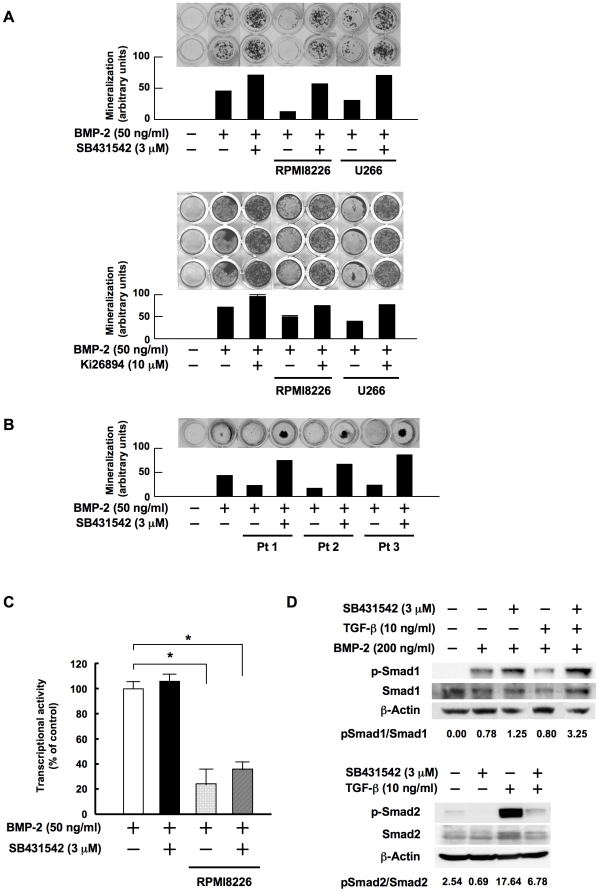
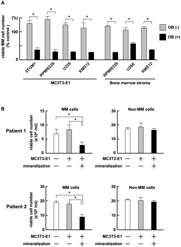
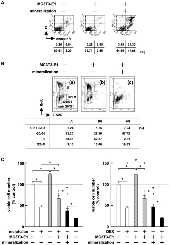
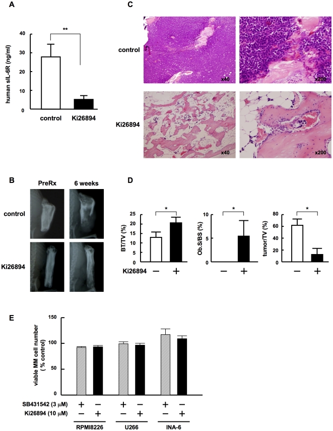
Methodology/principal findings: TGF-beta suppressed OB differentiation from bone marrow stromal cells and MC3T3-E1 preosteoblastic cells, and also inhibited adipogenesis from C3H10T1/2 immature mesenchymal cells, suggesting differentiation arrest by TGF-beta. Inhibitors for a TGF-beta type I receptor kinase, SB431542 and Ki26894, potently enhanced OB differentiation from bone marrow stromal cells as well as MC3T3-E1 cells. The TGF-beta inhibition was able to restore OB differentiation suppressed by MM cell conditioned medium as well as bone marrow plasma from MM patients. Interestingly, TGF-beta inhibition expedited OB differentiation in parallel with suppression of MM cell growth. The anti-MM activity was elaborated exclusively by terminally differentiated OBs, which potentiated the cytotoxic effects of melphalan and dexamethasone on MM cells. Furthermore, TGF-beta inhibition was able to suppress MM cell growth within the bone marrow while preventing bone destruction in MM-bearing animal models.

Conclusions/significance: The present study demonstrates that TGF-beta inhibition releases stromal cells from their differentiation arrest by MM and facilitates the formation of terminally differentiated OBs, and that terminally differentiated OBs inhibit MM cell growth and survival and enhance the susceptibility of MM cells to anti-MM agents to overcome the drug resistance mediated by stromal cells. Therefore, TGF-beta appears to be an important therapeutic target in MM bone lesions.


Conflict of interest statement

Competing Interests: The authors have declared that no competing interests exist.

Figures

Figure 1
Figure 1. TGF-β suppresses and TGF-β inhibitors enhance OB differentiation.
MC3T3-E1 cells (A) and primary bone marrow stromal cells (B) were cultured for 14 and 28 days, respectively, in 24-well culture plates in α-MEM containing 10% FBS supplemented with β-glycerophosphate and ascorbic acid (osteogenic medium). rhBMP-2, SB431542, Ki26894 and rhTGF-β were added at 50 ng/mL, 3 µM, 10 µM and 5 ng/mL to the indicated wells, respectively. After culturing, mineralized nodules were visualized by von Kossa staining. C. MC3T3-E1 cells were cultured for 21 days in osteogenic media in the absence of BMP-2. SB431542 was added at 3 µM to the indicated wells. Mineralized nodules were visualized by von Kossa staining. The data with mineralized nodule formation were quantified by densitometric analyses.
Figure 2
Figure 2. TGF-β suppresses adipogenic differentiation as well as OB differentiation from immature mesenchymal cells.
A. C3H10T1/2 immature mesenchymal cells were cultured in osteogenic medium for 7 days. rhBMP-2, SB431542 and rhTGF-β were added at 50 ng/mL, 3 µM and 5 ng/mL to the indicated wells, respectively. After culturing for 7days, the cells were stained with oil red O. Representative images of oil red O staining are shown. B. C3H10T1/2 cells were harvested after culturing for 2 days. rhBMP-2, SB431542 and rhTGF-β were added at 50 ng/mL, 3 µM and 5 ng/mL to the indicated wells, respectively. mRNA expression of markers for adipogenic and osteoblastic differentation were analyzed by RT-PCR. The primers used were as follows: Mouse adioponectin sense 5′-AGGGTGAGACAGGAGATGTTGGAA-3′ and antisense 5′-CAGAGGCCTGGTCCACATTCTTTT-3′. Mouse aP2 sense 5′-TCTCACCTGGAAGACAGCTCCTCCTCG-3′ and antisense 5′-TTCCATCCAGGCCTCTTCCTTTGGCTC-3′. Mouse ALP sense 5′-CACTCAGGGCAATGAGGTCACATC-3′ and antisense 5′-TTCAGTGCGGTTCCAGACATAGTG-3′.
Figure 3
Figure 3. TGF-β inhibition restores OB differentiation and potentiates Smad1 phosphorylation by BMP-2.
A. MC3T3-E1 cells were cultured for 14 days in osteogenic media in the absence or presence of conditioned media from RPMI8226 and U266 cells at 20%. SB431542 and Ki26894 were added at 3 and 10 µM to the indicated wells, respectively. B. MC3T3-E1 cells were cultured for 14 days in osteogenic media in the absence or presence of bone marrow plasma from patients with MM at 5%. SB431542 was added at 3 µM to the indicated wells. C. A chimeric firefly luciferase reporter plasmid (Topflash) and a renilla luciferase reporter plasmid (pRL-TK) were cotransfected into MC3T3-E1 cells as described in Materials and methods. The MC3T3-E1 cells were cultured in osteogenic media with rhBMP-2 at 50 ng/ml in the absence or presence of conditioned medium from RPMI8226 cells at 10%. SB431542 was added at 3 µM to the indicated wells. After 12 hours of incubation luiferase reporter activity was measured as renilla luiferase activity. Data were expressed as means +/− SD for six independent experiments. D. MC3T3-E1 cells were cultured in α-MEM with 1% FBS for 24 hours in 10-cm dishes at 60% confluence. SB431542 (3 µM), rhTGF-β (10 ng/mL), and the two in combination were added to the indicated dishes and the cells were cultured for another 48 hours, after which rhBMP-2 was added at 200 ng/ml (upper). SB431542 (3 µM), rhTGF-β (10 ng/mL), and the two in combination were added to the indicated dishes (lower). Following incubation for 30 minutes, cell lysates were collected. The protein levels of phosphorylated Smad1, Smad1, phosphorylated Smad2 and Smad2 were estimated by Western blotting. β-actin was used as a protein loading control.
Figure 4
Figure 4. Terminally differentiated OBs suppress MM cell growth.
A. MC3T3-E1 cells and primary bone marrow stromal cells isolated from a patient with MM were cultured in 24-well culture plates in α-MEM containing 10% FBS with or without β-glycerophosphate, ascorbic acid and 50 ng/mL rhBMP-2 as described in “Materials and methods”. After forming mineralized nodules in the osteogenic cultures with rhBMP-2, the cells were washed to remove rhBMP-2. MM cell lines, 5TGM1, RPMI8226, U266 and KMS-12 at 5×10⁴/mL, were cultured alone as a control or cocultured in quadruplicate with thus treated MC3T3-E1 cells or primary bone marrow stromal cells with or without inducing terminal OB differentiation. After culturing for 3 days, MM cells were harvested, and viable MM cell numbers were counted. Percent changes from the control are shown. Results are expressed as means +/− SD. *, <0.05. B. CD138-positive MM cells and CD138-negative non-MM bone marrow cells were immunomagnetically isolated from bone marrow aspirates of MM patients. The cells were cultured alone or co-cultured in triplicate with MC3T3-E1 cells with or without mineralization. After culturing for 3 days, the MM and non-MM bone marrow cells were harvested and viable cell numbers were counted. Data were expressed as means +/− SD. *, p<0.05.
Figure 5
Figure 5. Terminally differentiated OBs induce MM cell apoptosis with G1 arrest.
A. 5TGM1 cells (5×10⁴/mL) were cultured alone or co-cultured with MC3T3-E1 cells with or without mineralized nodules. After culturing for 2 days, 5TGM1 cells were collected, stained with FITC-labeled annexin V and PI, and analyzed by flow cytometry. B. 5TGM1 cells were cultured alone, or cocultured with MC3T3-E1 cells with or without mineralized nodules for 2 days. 5TGM1 cells were analyzed by flow cytometry with dual staining of BrdU and 7-AAD as described in “Materials and methods”. Cells were gated corresponding to cell cycle distribution. C. RPMI8226 cells were cultured alone or cocultured with MC3T3-E1 cells with or without mineralization in quadruplicate for 3 days in the absence or presence of melphalan at 20 µM (left) or dexamethasone at 10 µM (right). Viable RPMI8226 cells were counted.
Figure 6
Figure 6. TGF-β inhibition facilitates terminal OB maturation to suppress MM growth and survival at earlier time points.
A. MC3T3-E1 cells were cultured in osteogenic media for the indicated periods. rhBMP-2, SB431542 and rhTGF-β were added at 50 ng/mL, 3 µM and 5 ng/mL to the indicated wells, respectively. After culturing, mineralized nodules were visualized by von Kossa staining. B. MC3T3-E1 cells were cultured in osteogenic media for induction of OB differentiation. rhBMP-2 and SB431542 were added at 50 ng/mL and 3 µM to the indicated wells, respectively. After culturing for 3, 6 and 9 days, the cells were washed, and subjected to the measurement of ALP activity (top panels) and von Kossa staining for assessing mineralized nodule formation (middle panels). The cells were also cocultured in quadruplicate with 5TGM1 cells for 3 days. Viable 5TGM1 cells were counted. Percent changes from the baseline are shown (bottom panels). Data are expressed as means +/− SD. *, p<0.05.
Figure 7
Figure 7. TGF-β inhibition suppresses MM cell growth in bone as well as bone destruction.
INA6-bearing SCID-rab mice were given food containing either the TGF-beta type I receptor kinase inhibitor Ki26894 or a vehicle control. A. Mouse serum samples were collected 6 weeks after the inoculation of INA6 cells and serum levels of the soluble human IL-6 receptor were measured as described in Materials and methods. **, <0.01. B. X-ray photographs of the rabbit bones were taken before and 6 weeks after the treatment. Representative images are shown. C. At 6 weeks after the treatment, the rabbit bones were removed. The samples were sectioned and stained with H&E. Representative specimens of control and treated rabbit bones are shown in the upper and lower panels, respectively. Original magnifications were ×40 and ×200 as indicated. D. Histomorphometrical analyses of the implanted rabbit bone sections. BV/ TV, Ob.S/BS and tumor/TV were measured in decalcified implanted rabbit bone sections stained with H&E as described in Materials and Methods. Data are expressed as means +/− SD. *, p<0.05. E. MM cell lines, INA6, RPMI8226, U266 and KMS-12 at 5×10⁴/mL, were cultured in the absence or presence of Ki26894 (10 µM) or SB431542 (3 µM). After culturing for 3 days, viable MM cell numbers were counted. Percent changes from the control are shown. Results are expressed as means +/− SD.

References

    1. Abe M, Hiura K, Wilde J, Moriyama K, Hashimoto T, et al. Role for macrophage inflammatory protein (MIP)-1alpha and MIP-1beta in the development of osteolytic lesions in multiple myeloma. Blood. 2002;100:2195–2202. - PubMed
    1. Hashimoto T, Abe M, Oshima T, Shibata H, Ozaki S, et al. Ability of myeloma cells to secrete macrophage inflammatory protein (MIP)-1alpha and MIP-1beta correlates with lytic bone lesions in patients with multiple myeloma. Br J Haematol. 2004;125:38–41. - PubMed
    1. Choi SJ, Oba Y, Gazitt Y, Alsina M, Cruz J, et al. Antisense inhibition of macrophage inflammatory protein 1-alpha blocks bone destruction in a model of myeloma bone disease. J Clin Invest. 2001;108:1833–1841. - PMC - PubMed
    1. Abe M, Hiura K, Ozaki S, Kido S, Matsumoto T. Vicious cycle between myeloma cell binding to bone marrow stromal cells via VLA-4-VCAM-1 adhesion and macrophage inflammatory protein-1alpha and MIP-1beta production. J Bone Miner Metab. 2009;27:16–23. - PubMed
    1. Oshima T, Abe M, Asano J, Hara T, Kitazoe K, et al. Myeloma cells suppress bone formation by secreting a soluble Wnt inhibitor, sFRP-2. Blood. 2005;106:3160–3165. - PubMed

Publication types

MeSH terms