. 2010 Mar 26;5(3):e9863.
doi: 10.1371/journal.pone.0009863.

An expanded self-antigen peptidome is carried by the human lymph as compared to the plasma

Affiliations

An expanded self-antigen peptidome is carried by the human lymph as compared to the plasma

Cristina C Clement et al. PLoS One. .

Abstract

Background: The pre-nodal afferent lymph is the fluid which directly derives from the extracellular milieu from every parenchymal organ and, as it continues to circulate between the cells, it collects products deriving from the organ metabolism/catabolism. A comprehensive qualitative and quantitative investigation of the self-antigenic repertoire transported by the human lymph is still missing.

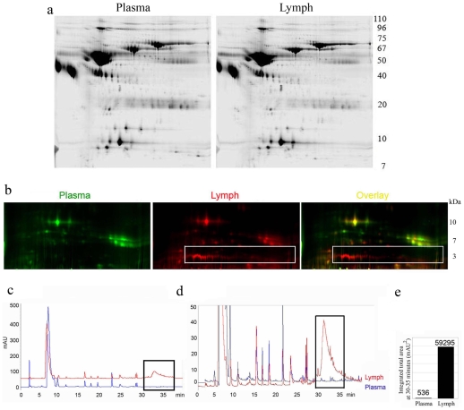
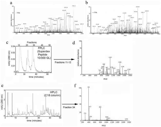
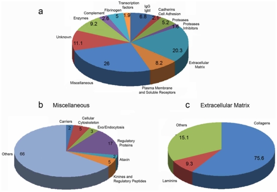
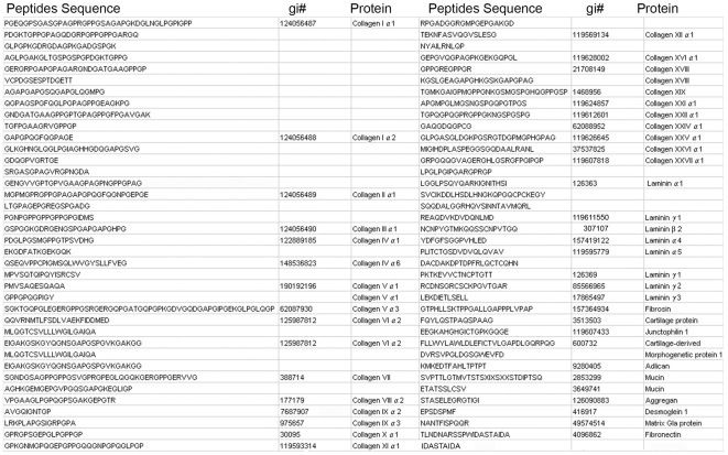
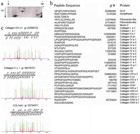
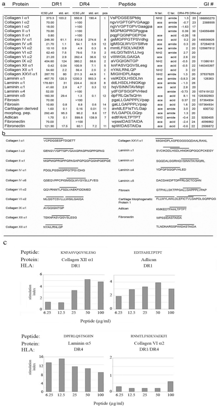
Methodology/principal findings: A major difference between lymph and plasma could be visualized by FPLC and 2D gel in the amount of low molecular weight products corresponding to peptide fragments. Naturally processed peptides in normal pre-nodal human lymph were then fractionated by HPLC and characterized by multidimensional mass spectrometry. Analysis of more then 300 sequences identified self-peptides derived from both intracellular and extracellular proteins revealing the variety of catabolic products transported by human lymph. Quantitative analysis established that at least some of these peptides are present in the circulating lymph in nanomolar concentration.

Conclusions/significance: The peptidome, generated by physiological tissue catabolism and transported by the pre-nodal lymph, is in addition to the self-peptidome generated in endosomal compartment. Unlike self antigen processed by local or nodal APC, which mostly produce epitopes constrained by the endosomal processing activity, self antigens present in the lymph could derived from a wider variety of processing pathways; including caspases, involved in cellular apoptosis, and ADAM and other metalloproteinases involved in surface receptor editing, cytokines processing and matrix remodeling. Altogether, expanding the tissue-specific self-repertoire available for the maintenance of immunological tolerance.


Conflict of interest statement

Competing Interests: The authors have declared that no competing interests exist.

Figures

Figure 1
Figure 1. Comparative peptidomic profile of human lymph and plasma samples.
a) Comparative 2D-gel analysis of human lymph and plasma proteins (2CyDye). b) Fluorescence (2CyDye) comparative 2D-gel analysis of human lymph and plasma low molecular weight products (1–10 kDa). One out of 6 silver staining gels is shown c) Overlays of reverse phase HPLC profile of lymph and plasma low MW filtrates (<5000 Da). Samples from 18 subjects were pooled. d) is a 10 times magnification of c. Boxed areas indicate where major differences between the two samples are observed. One out of four reverse phase HPLC analysis is shown. e) Bar graph representing the integration number calculated from the curves in the boxed area for both the lymph and the plasma pooled samples.
Figure 2
Figure 2. MS peptidomic profile of the human lymph.
a and b) Mass spectroscopy scan (200–2000 m/z) of low MW (5000 Da) filtrate from lymph samples obtained before or after elution in 0.1% TFA. c) Elution profile of the low MW filtrate (<5000 Da) collected from the lymph pooled samples run through a superdex peptide 10/300 GL column d) MS scan of eluted fractions 11–12. e) Representative elution profile of the low MW filtrate (<5000 Da) collected from the lymph pooled samples run through a C18 column f) Representative full MS scan of eluted fraction 34.
Figure 3
Figure 3. Schematic representation of the MS/MS peptidomic profile of the human lymph.
a, b and c) Pie chart representation of self-peptides (<5000 Da) sequenced from the human lymph. Data are compiled from 4 independent MS/MS analysis.
Figure 4
Figure 4. Amino acid sequence of self-peptides derived from processing of extracellular matrix proteins found in the human lymph.
MS/MS sequencing analysis of peptides found in the human lymph, which derive from processing of extracellular matrix proteins.
Figure 5
Figure 5. Quantification of 2D gel eluted peptide.
a) Silver staining of an SDS-PAGE run with 20 microgram of immunoglobulin and albumin depleted lymph. b) Sequence of some of the peptides retrieved from each of the analyzed spot. c) MS/MS fragmentation profile and respective sequence of the three peptides eluted from spot number 6. Green dotted lines identified assigned fragmentation picks. Total peptide concentration eluted from spot number 6 calculated at 230 OD was 0.22 µM.
Figure 6
Figure 6. HLA-DR1 and HLA-DR4 binding affinity and T cell proliferative response to some of the lymph sequenced peptides.
a) IC50, as determined by competitive fluorescence polarization assay, for short peptides derived from fragments identified in lymph. Peptides were selected based on predicted binding of nonameric binding frames (capital letters) to HLA-DR1 or HLA-DR4. Peptides were synthesized with N-terminal acetyl and C-terminal amide groups unless the peptide termini were coincident with an end of the sequenced fragment, in which case the native terminus was used b) Position of the assayed peptides in the sequenced fragments indicated by bars above the sequence. c) Proliferative responses (calculated by thymidine incorporation assay) in lymph nodes from peptide immunized mice. One out of four proliferation assay is shown for each peptide.

Similar articles

Cited by

References

    1. Savina A, Amigorena S. Phagocytosis and antigen presentation in dendritic cells. Immunol Rev. 2007;219:143–156. - PubMed
    1. Vermaelen KY, Carro-Muino I, Lambrecht BN, Pauwels RA. Specific migratory dendritic cells rapidly transport antigen from the airways to the thoracic lymph nodes. J Exp Med. 2001;193:51–60. - PMC - PubMed
    1. Pabst O, Bernhardt G, Forster R. The impact of cell-bound antigen transport on mucosal tolerance induction. J Leukoc Biol. 2007;82:795–800. - PubMed
    1. Jahnsen, FL, Strickland DH, Thomas JA, Tobagus IT, Napoli S, et al. Accelerated antigen sampling and transport by airway mucosal dendritic cells following inhalation of a bacterial stimulus. J Immunol. 2006;177:5861–5867. - PubMed
    1. Hunger RE, Yawalkar N, Braathen LR, Brand CU. CD1a-positive dendritic cells transport the antigen DNCB intracellularly from the skin to the regional lymph nodes in the induction phase of allergic contact dermatitis. Arch Dermatol Res. 2001;293:420–426. - PubMed

Publication types