. 2010 Mar 25;5(3):e9898.
doi: 10.1371/journal.pone.0009898.

Specific functions of BIG1 and BIG2 in endomembrane organization

Affiliations

Specific functions of BIG1 and BIG2 in endomembrane organization

Frédéric Boal et al. PLoS One. .

Abstract

Background: Transport of molecules from one subcellular compartment to another involves the recruitment of cytosolic coat protein complexes to a donor membrane to concentrate cargo, deform the membrane and ultimately to form an independent carrier. Small-GTP-binding proteins of the Arf family are central to many membrane trafficking events. Arfs are activated by guanine nucleotide exchange factors (GEFs) which results in their recruitment to membranes and subsequent engagement with Arf-effectors, many of which are coat proteins. Among the human BFA-sensitive large Arf-GEFs, the function of the two closely related BIG1 and BIG2 is still not clear, and recent studies have raised the question of functional redundancy between the two proteins.

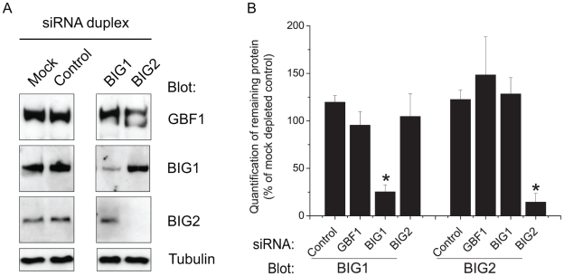
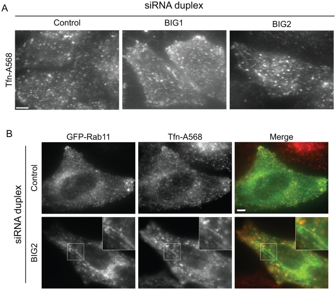
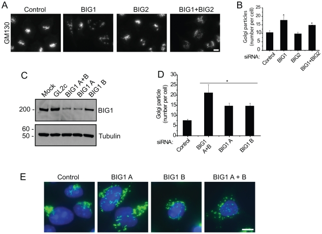
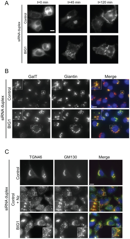
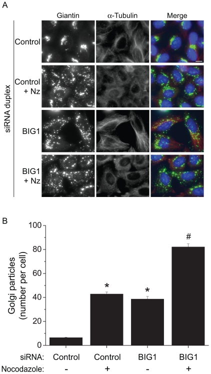
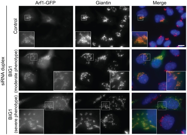
Methodology/principal findings: Here we have used small-interfering RNA on human cells and a combination of fixed and live-cell imaging to investigate the differential functions of BIG1 and BIG2 in endomembrane organization and function. Importantly, in this direct comparative study, we show discrete functions for BIG1 and BIG2. Our results show that depletion of BIG2 but not of BIG1 induces a tubulation of the recycling endosomal compartment, consistent with a specific role for BIG2 here. In contrast, suppression of BIG1 induces the formation of Golgi mini-stacks still polarized and functional in terms of cargo export.

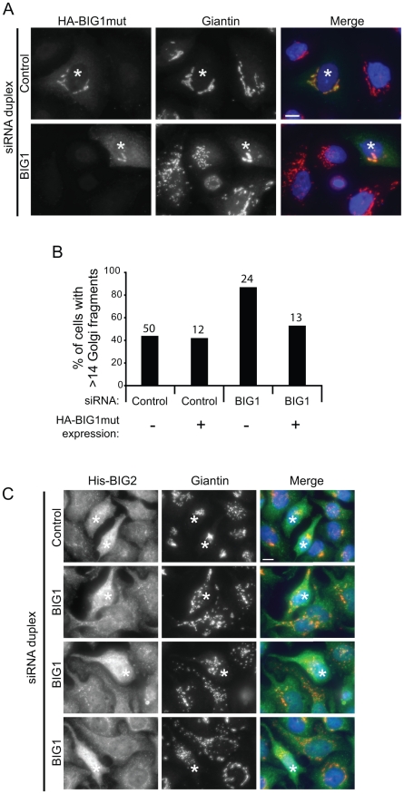
Conclusions: A key finding from our work is that suppression of BIG1 expression results in a fragmentation of the Golgi apparatus. Our data indicate that the human BFA-sensitive large Arf-GEFs have non-redundant functions in cell organization and membrane trafficking. BIG1 is required to maintain the normal morphology of the Golgi; BIG2 is important for endosomal compartment integrity and cannot replace the function of BIG1 in Golgi organization.


Conflict of interest statement

Competing Interests: The authors have declared that no competing interests exist.

Figures

Figure 1
Figure 1. Depletion of human large-GEFs using siRNA.
A: HeLa cells were transfected without siRNA (Mock), with non-targeting siRNA duplexes (Control), or with siRNA targeting indicated GEF (GBF1, BIG1, BIG2). Cell lysates were then immunoblotted for antibodies indicated on the left (Blot). Arrowhead points to the specific band for BIG1. B: Western-blots were quantified by densitometry and protein contents are expressed as percentage of mock-transfected cells. Results are presented as mean +/− s.e.m., n = 3–4, asterisk means p<0.05 as compared to cells transfected with a non-targeted siRNA (Control). Molecular-mass markers are shown in kDa in all figures.
Figure 2
Figure 2. Depletion of BIG2 induces the tubulation of the endosomal compartment.
A: HeLa cells depleted using siRNA duplexes as indicated were loaded with fluorescent transferrin (Tfn-A568) for 30 minutes prior to live-cell imaging. Movie S1 shows the stability of the BIG2-depletion induced tubules. B: Tfn-A568 loaded cells were co-labelled using GFP-Rab11. Single optical z-sections are shown resulting in the presence of some out-of-focus light, and panels below show enlarged regions. Bar = 10 µm in all figures otherwise stated.
Figure 3
Figure 3. BIG1 suppression induces Golgi fragmentation.
A: GM130 labelling reveals fragmentation of the Golgi apparatus following suppression of BIG1 or BIG1 and BIG2 together, but control siRNA duplexes or duplexes targeting BIG2 alone do not induce this phenotype. B: After applying the same threshold to all images, the number of Golgi fluorescent particles per cell was determined as described in “Materials and Methods”. Results are presented as mean +/− s.e.m., asterisk means p<0.05 as compared to Control-depleted cells, n = 3. C: BIG1 suppression using individual siRNA duplexes was monitored by immunoblotting. Tubulin is used as a loading control. D: Quantification of Golgi-fragmentation done as in B shows that both individual siRNA give a similar phenotype. E: Cells depleted of BIG1 using individual or pooled duplexes were labelled with antibodies against the Golgi matrix protein giantin (in green) and nuclei are counterstained with DAPI (in blue).
Figure 4
Figure 4. Rescue of Golgi fragmentation in BIG1-suppressed cells.
A: Control cells, or those depleted of BIG1 expression were transfected to express a HA-tagged mutant BIG1 (HA-BIG1mut) that is resistant to the siRNA. Cells were processed for immunofluorescence using anti-HA (green) and anti-giantin (red) antibodies. Asterisks indicate transfected cells, showing the rescued phenotype in BIG1-depleted cells. B: Quantification of the rescue phenotype. Cells were transfected with indicated siRNA and subsequently transfected to express HA-BIG1mut. The number of Golgi particles per cell was counted as in Fig. 3B. Data are pooled from two independent experiments. The numbers on top of the bars indicate the number of cells counted for each condition. C: BIG2 expression cannot rescue the fragmentation of the Golgi induced by BIG1-depletion. Control cells or those depleted of BIG1 expression were transfected to express a His-tagged BIG2 (His-BIG2). Cells were labelled using anti-BIG2 (green) and anti-giantin (red) antibodies. Asterisks indicate transfected cells, showing that overexpression of BIG2 does not rescued the phenotype in BIG1-depleted cells. Three different examples are shown for BIG1-depleted cells.
Figure 5
Figure 5. The fragmented Golgi induced by BIG1 suppression remains functional.
A: Cells were infected with adenovirus to express tsO45-G-YFP at 39.5°C. Cells were then shifted to 32°C to allow the trafficking of the viral protein for 0, 45, or 120 minutes as indicated. Trafficking of the viral protein is indistinguishable between control and BIG1 depleted cells. Note the fragmentation of the Golgi in BIG1 depleted cells confirming the efficacy of BIG1 suppression. B: The fragmented Golgi in BIG1-depleted cells is still populated with endogenous galactosyltransferase (GalT). Depleted HeLa cells were processed for immunofluorescence with anti-GalT (green in merge) and anti-giantin (red) antibodies. C: Scattered Golgi elements induced by BIG1-depletion retain their cis-trans polarity. HeLa cells transfected with siRNA duplexes as indicated were incubated with nocodazole (Nz) to depolymerize the microtubules, methanol-fixed, and processed for immunofluorescence with anti-TGN46 (green in merge) and anti-GM130 (red) antibodies.
Figure 6
Figure 6. The Golgi mini-stacks in BIG1-depleted cells are still coordinated on microtubules.
A: HeLa transfected for indicated siRNA were treated or not with nocodazole (Nz) and stained for giantin and α-tubulin. B: Quantification of Golgi-particles was done as in previous figures. A higher fragmentation of the Golgi can be achieved by treating BIG1-depleted cells with nocodazole, suggesting that the Golgi mini-stacks are still coordinated on microtubules. Asterisks mean p<0.05 as compared to control untreated cells, # means p<0.05 as compared to nocodazole-treated BIG1-deplepted cells.
Figure 7
Figure 7. Arf1 is still recruited to Golgi mini-stacks in BIG1-depleted cells.
HeLa cells depleted of BIG1 and transiently expressing Arf1-GFP were processed for immunofluorescence with an anti-giantin (red) antibody.

Similar articles

Cited by

References

    1. Bonifacino JS, Glick BS. The mechanisms of vesicle budding and fusion. Cell. 2004;116:153–166. - PubMed
    1. Duden R. ER-to-Golgi transport: COP I and COP II function. Molecular Membrane Biology. 2003;20:197–207. - PubMed
    1. Robinson MS. Adaptable adaptors for coated vesicles. Trends Cell Biol. 2004;14:167–174. - PubMed
    1. Kuge O, Dascher C, Orci L, Rowe T, Amherdt M, et al. Sar1 promotes vesicle budding from the endoplasmic reticulum but not Golgi compartments. Journal of Cell Biology. 1994;125:51–65. - PMC - PubMed
    1. Gillingham AK, Munro S. The small g proteins of the arf family and their regulators. Annu Rev Cell Dev Biol. 2007;23:579–611. - PubMed

Publication types

MeSH terms