

A 'brain tumor' in an intravenous drug abuser
- PMID: 20360890
- PMCID: PMC2840556
- DOI: 10.2147/ijgm.s4826
A 'brain tumor' in an intravenous drug abuser
Abstract
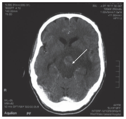
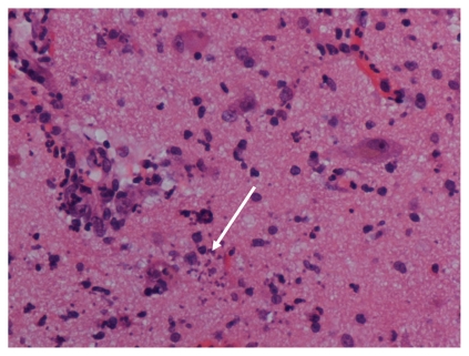
A male intravenous drug abuser who was infected with hepatitis B and C, presented with a slowly progressive hemiplegia. Contrast enhanced computerized tomography of the head showed a solitary ring-enhanced mass with surrounding edema. Clinically brain tumor was suspected but a brain biopsy confirmed cerebral toxoplasmosis. An HIV test was not considered until the result of brain biopsy. He also had lymphopenia and positive serum toxoplasma antibody. His subsequent HIV test was positive. He deteriorated after a brain biopsy. Empirical antitoxoplasma treatment is recommended in HIV-positive patients with ring-enhanced lesions with surrounding edema and with positive toxoplasma serology. Cerebral toxoplasmosis is still the commonest cerebral opportunistic infection in HIV-infected patients even though the incidence has declined with the use of antiretroviral therapy. It is often diagnosed in those patients as an initial presentation of HIV infection or in those who failed to attend for disease monitoring. Clinical features and differential diagnosis of cerebral toxoplasmosis in immunocompromised patients are discussed.
Keywords: HIV/AIDS; brain tumor; cerebral toxoplasmosis; intravenous drug abuser; primary brain lymphoma.
Figures
Similar articles
-
Multiple intracerebral lesions in a young male.Afr Health Sci. 2015 Sep;15(3):1045-8. doi: 10.4314/ahs.v15i3.47. Afr Health Sci. 2015. PMID: 26958001 Free PMC article.
-
SPECT thallium-201 combined with Toxoplasma serology for the presumptive diagnosis of focal central nervous system mass lesions in patients with AIDS.J Infect. 2000 May;40(3):274-81. doi: 10.1053/jinf.2000.0664. J Infect. 2000. PMID: 10908023
-
IgG4 specific to Toxoplasma gondii excretory/secretory antigens in serum and/or cerebrospinal fluid support the cerebral toxoplasmosis diagnosis in HIV-infected patients.J Immunol Methods. 2013 Sep 30;395(1-2):21-8. doi: 10.1016/j.jim.2013.06.005. Epub 2013 Jun 26. J Immunol Methods. 2013. PMID: 23811152
-
Cerebral toxoplasmosis with multiple hemorrhage lesions in an HIV infected patient: A case report and literature review.Parasitol Int. 2021 Apr;81:102280. doi: 10.1016/j.parint.2020.102280. Epub 2021 Jan 2. Parasitol Int. 2021. PMID: 33401016 Review.
-
Spinal cord toxoplasmosis in human immunodeficiency virus infection/acquired immunodeficiency syndrome.Infect Dis (Lond). 2015 May;47(5):277-82. doi: 10.3109/00365548.2014.993421. Infect Dis (Lond). 2015. PMID: 25835092 Review.
Cited by
-
An unusual cause of isolated secondary ovarian failure due to cerebral toxoplasmosis in an African woman with AIDS.BMJ Case Rep. 2017 Aug 7;2017:bcr2017221337. doi: 10.1136/bcr-2017-221337. BMJ Case Rep. 2017. PMID: 28784911 Free PMC article.
-
Multiple intracerebral lesions in a young male.Afr Health Sci. 2015 Sep;15(3):1045-8. doi: 10.4314/ahs.v15i3.47. Afr Health Sci. 2015. PMID: 26958001 Free PMC article.
References
-
- Navia BA, Petito CK, Gold JW, Cho ES, Jordan BD, Price RW. Cerebral toxoplasmosis complicating the acquired immune deficiency syndrome: clinical and neuropathological findings in 27 patients. Ann Neurol. 1986;19:224–238. - PubMed
-
- Piccolo I, Causarano R, Sterzi R, et al. Chorea in patients with AIDS. Acta Neurol Scand. 1999;100:332–336. - PubMed
-
- Rebaud C, May T, Amiel C, et al. Extracerebral toxoplasmosis in patients infected with HIV. A French National Survey. Medicine (Baltimore) 1994;73:306–314. - PubMed
-
- Ammassari A, Cingolani A, Pezzotti P, et al. AIDS-related focal brain lesions in the era of highly active antiretroviral therapy. Neurology. 2000;55:1194–1200. - PubMed
-
- Skiest DJ, Erdman W, Chang WE, Oz OK, Ware A, Fleckenstein J. SPECT thallium-201 combined with Toxoplasma serology for the presumptive diagnosis of focal central nervous system mass lesions in patients with AIDS. J Infect. 2000;40:274–281. - PubMed
Publication types
LinkOut - more resources
Full Text Sources
