. 2010 Mar 26;6(3):e1000836.
doi: 10.1371/journal.ppat.1000836.

T cell-dependence of Lassa fever pathogenesis

Affiliations

T cell-dependence of Lassa fever pathogenesis

Lukas Flatz et al. PLoS Pathog. .

Abstract

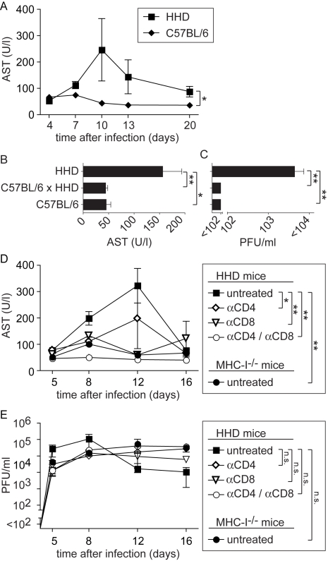
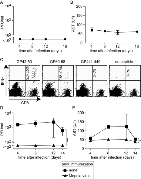
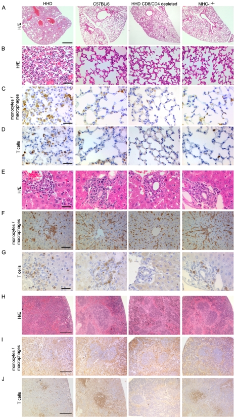
Lassa virus (LASV), the causative agent of Lassa fever (LF), is endemic in West Africa, accounting for substantial morbidity and mortality. In spite of ongoing research efforts, LF pathogenesis and mechanisms of LASV immune control remain poorly understood. While normal laboratory mice are resistant to LASV, we report that mice expressing humanized instead of murine MHC class I (MHC-I) failed to control LASV infection and develop severe LF. Infection of MHC-I knockout mice confirmed a key role for MHC-I-restricted T cell responses in controlling LASV. Intriguingly we found that T cell depletion in LASV-infected HHD mice prevented disease, irrespective of high-level viremia. Widespread activation of monocyte/macrophage lineage cells, manifest through inducible NO synthase expression, and elevated IL-12p40 serum levels indicated a systemic inflammatory condition. The absence of extensive monocyte/macrophage activation in T cell-depleted mice suggested that T cell responses contribute to deleterious innate inflammatory reactions and LF pathogenesis. Our observations in mice indicate a dual role for T cells, not only protecting from LASV, but also enhancing LF pathogenesis. The possibility of T cell-driven enhancement and immunopathogenesis should be given consideration in future LF vaccine development.


Conflict of interest statement

The authors have declared that no competing interests exist.

Figures

Figure 1
Figure 1. MHC-I but not MHC-II determines efficient LASV control.
Mice of the indicated genotypes were infected with LASV. A, B: Blood samples were collected over time to determine viremia. A: The 2.5 hrs, 2 day and 4 day time points (left part of panel A) were determined in one experiment, and day 7, 10, 13 and 20 (right part of panel A) were determined in a second one. C: Mice were sacrificed on day 2, 4 and 8 after LASV infection to measure viral titers in organs. Axis breaks indicate the technical detection limit. Symbols and bars represent the mean±SEM of three to five mice per group.
Figure 2
Figure 2. MHC-I- and T cell-dependent AST elevation suggest T cell-dependent immunopathogenesis of LF.
A: C57BL/6 and HHD mice were infected with LASV. AST activity was determined in serum samples collected over time. B, C: In a separate experiment comprising C57BL/6, C57BL/6 x HHD F1 and HHD mice, serum AST activity (B) and viremia (C) were measured on day 8 after LASV infection. D, E: HHD mice were either left untreated, were depleted of CD8+ T cells or of CD4+ T cells or were depleted of both populations. All groups of HHD mice and MHC-I-/- mice were subsequently infected with LASV. Serum AST activity (D) and viremia (E) was measured over time. Symbols and bars represent the mean±SEM of three to five mice per group and time point.
Figure 3
Figure 3. Mopeia virus (MV) infection is apathogenic in HHD mice but elicits LASV-specific HLA-A2.1-restricted CD8+ T cells and protects against subsequent LASV challenge.
A, B: HHD mice were infected with MV. Serum and blood samples were collected at the indicated time points for measuring viremia (A) and AST (B), respectively. C: Epitope-specific CD8+ T cells in the spleen of MV-infected HHD mice were measured on day 8 (naive controls did not display detectable responses, data not shown). The percent range of IFNγ-producing CD8+ T cells is indicated in the upper right quadrant. D, E: HHD mice, either naive or infected with MV two weeks previously, were challenged with LASV. Viremia (D) and serum AST activity (E) were measured at the indicated time points. Symbols represent the mean ±SEM of four to five mice per group and time point.
Figure 4
Figure 4. T cell- and MHC-I-dependent alterations in the tissues of LASV-infected mice.
HHD, C57BL/6, HHD mice depleted of CD8+ and CD4+ T cells, and MHC-I-/- mice were infected with LASV. Eight days later, lung (A-D), liver (E-G) and spleen (H-J) tissue was processed for histological analysis. H/E (A, B, E, H) or immunohistochemical staining of monocytes/macrophages (Iba-1; C, F, I) and T cells (CD3; D, G, J) are shown. Note the collapsed alveolar space, rounded and irregularly distributed monocytes/macrophages in liver and disruption of the splenic microarchitecture in LASV-infected HHD mice. These changes are less severe or largely restored in C57/BL/6 and CD8/CD4-depleted HHD mice, respectively, whereas MHC-I-/- mice display an intermediate picture. Magnification bars indicate 1000 µm (A), 100 µm (B), 50 µm (C, D, F), 30 µm E,G) and 200 µm (H-J), respectively.
Figure 5
Figure 5. Monocytes/macrophages represent a major target of LASV and produce inflammatory mediators in a T cell-dependent fashion.
A, B: HHD, C57BL/6, HHD mice depleted of CD8+ and CD4+ T cells, and MHC-I-/- mice were infected with LASV. A: Eight days later, liver tissue was processed for histological analysis of iNOS expression. B. Serum samples were collected over time and IL-12p40 was determined by ELISA. C: Immunohistochemical staining of LASV-NP in lung, liver and spleen of HHD mice on day 8 after infection. D: Illustrative examples for cellular colocalization of the monocyte/macrophage marker Iba-1 (red) and LASV-NP (green) in the liver. Magnification bars indicate 50 µm (A, C) and 10 µm (D), respectively. E: Schematic describing the postulated mechanism how T cells may enhance LF pathogenesis.

References

    1. Buckley SM, Casals J, Downs WG. Isolation and antigenic characterization of Lassa virus. Nature. 1970;227:174. - PubMed
    1. McCormick JB, Webb PA, Krebs JW, Johnson KM, Smith ES. A prospective study of the epidemiology and ecology of Lassa fever. J Infect Dis. 1987;155:437–444. - PubMed
    1. Haas WH, Breuer T, Pfaff G, Schmitz H, Kohler P, et al. Imported Lassa fever in Germany: surveillance and management of contact persons. Clin Infect Dis. 2003;36:1254–1258. - PubMed
    1. Borio L, Inglesby T, Peters CJ, Schmaljohn AL, Hughes JM, et al. Hemorrhagic fever viruses as biological weapons: medical and public health management. Jama. 2002;287:2391–2405. - PubMed
    1. Charrel RN, de Lamballerie X. Arenaviruses other than Lassa virus. Antiviral Res. 2003;57:89–100. - PubMed

Publication types

MeSH terms