. 2010 Mar 26;6(3):e1000829.
doi: 10.1371/journal.ppat.1000829.

An accessory to the 'Trinity': SR-As are essential pathogen sensors of extracellular dsRNA, mediating entry and leading to subsequent type I IFN responses

Affiliations

An accessory to the 'Trinity': SR-As are essential pathogen sensors of extracellular dsRNA, mediating entry and leading to subsequent type I IFN responses

Stephanie J DeWitte-Orr et al. PLoS Pathog. .

Abstract

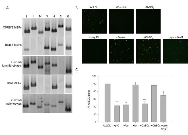
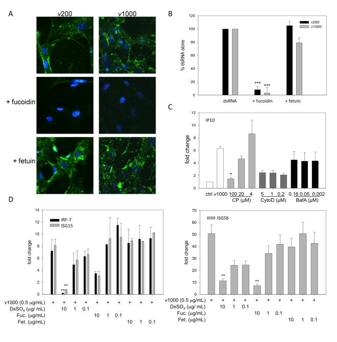
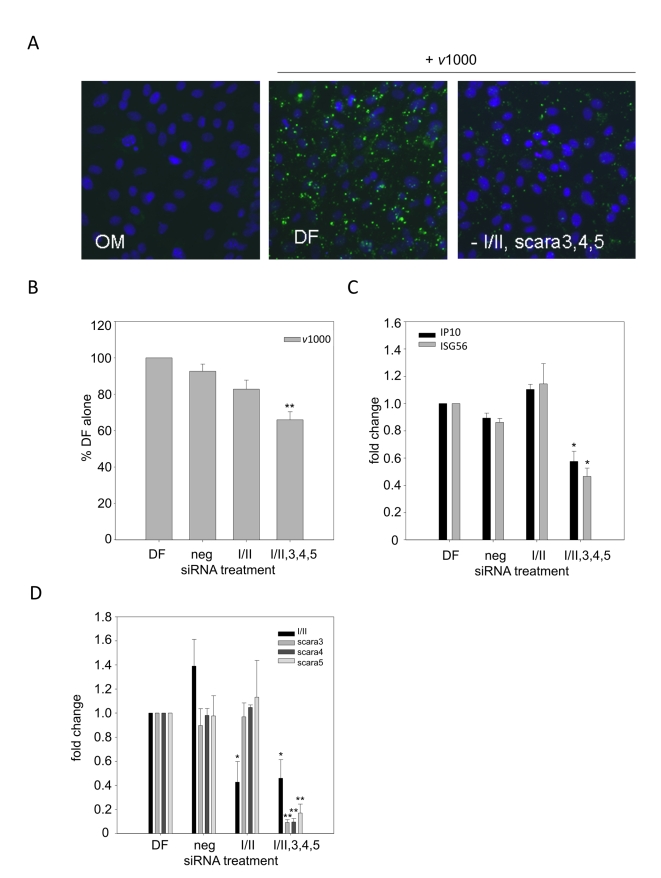
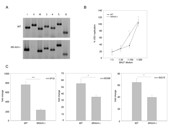
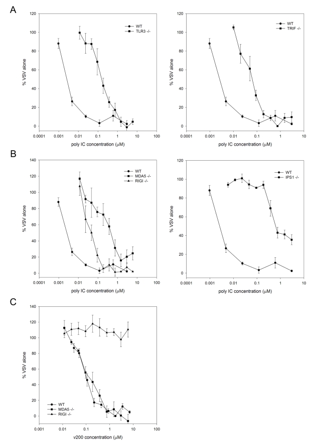
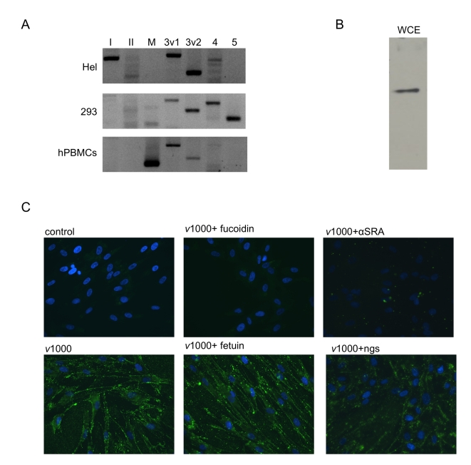
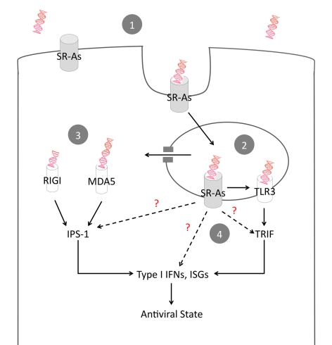
Extracellular RNA is becoming increasingly recognized as a signaling molecule. Virally derived double stranded (ds)RNA released into the extracellular space during virus induced cell lysis acts as a powerful inducer of classical type I interferon (IFN) responses; however, the receptor that mediates this response has not been identified. Class A scavenger receptors (SR-As) are likely candidates due to their cell surface expression and ability to bind nucleic acids. In this study, we investigated a possible role for SR-As in mediating type I IFN responses induced by extracellular dsRNA in fibroblasts, a predominant producer of IFNbeta. Fibroblasts were found to express functional SR-As, even SR-A species thought to be macrophage specific. SR-A specific competitive ligands significantly blocked extracellular dsRNA binding, entry and subsequent interferon stimulated gene (ISG) induction. Candidate SR-As were systematically investigated using RNAi and the most dramatic inhibition in responses was observed when all candidate SR-As were knocked down in unison. Partial inhibition of dsRNA induced antiviral responses was observed in vivo in SR-AI/II(-/-) mice compared with WT controls. The role of SR-As in mediating extracellular dsRNA entry and subsequent induced antiviral responses was observed in both murine and human fibroblasts. SR-As appear to function as 'carriers', facilitating dsRNA entry and delivery to the established dsRNA sensing receptors, specifically TLR3, RIGI and MDA-5. Identifying SR-As as gatekeepers of the cell, mediating innate antiviral responses, represents a novel function for this receptor family and provides insight into how cells recognize danger signals associated with lytic virus infections. Furthermore, the implications of a cell surface receptor capable of recognizing extracellular RNA may exceed beyond viral immunity to mediating other important innate immune functions.


Conflict of interest statement

The authors have declared that no competing interests exist.

Figures

Figure 1
Figure 1. Primary murine fibroblasts express functional SR-As.
(A) SR-A transcripts were detected using RT-PCR and primers specific to SR-AI (lane I), SR-AII (II), MARCO (M), SCARA3 (3), SCARA4 (4), and SCARA5 (5) in murine embryonic fibroblasts (MEFs) derived from C57Bl/6 or balb-c mice, the macrophage-like cell line RAW 264.7, and primary lung fibroblasts and splenocytes derived from C57Bl/6 mice. GAPDH was used as an internal control for all cell types (G). (B) Cell associated AcLDL was observed by live-cell fluorescence microscopy in MEFs derived from C57Bl/6 mice treated with Alexafluor 488 labeled AcLDL for 1h (2.5 µg/mL), in the presence or absence of SR-A competitive ligands (fucoidin, DxSO4), their corresponding non-competitive counterparts (fetuin, ChSO4) or nucleic acids (poly IC, poly dA:dT) all at 100 µg/mL. Magnification 200X. (C) AcLDL entry was measured using a fluorescence plate reader assay. Cells were treated with Alexafluor 488 labeled AcLDL (2.5µg/ mL) for 1h in the presence of poly IC (pIC), fucoidin (fuc), fetuin (fet), DxSO4, ChSO4 or poly dA:dT (100 µg/mL). Cells treated with AcLDL alone were set at 100% fluorescence. These data include three independent experiments ± SEM. Statistical analysis was performed by a one-way ANOVA with Tukey post test (* p<0.05, *** p<0.001).
Figure 2
Figure 2. SR-As mediate viral dsRNA binding, entry and resulting ISG induction in MEFs using a mechanism dependent on clathrin-mediated endocytosis.
(A) Cellular binding of viral dsRNA was observed by fluorescence microscopy in MEFs derived from C57Bl/6 mice treated with either 200 bp or 1000 bp Alexafluor 488 labeled viral dsRNA (v200 or v1000 respectively, both at 1 µg/mL) for 1h, in the presence or absence of fucoidin or fetuin (both at 100 µg/mL). Cells were fixed and nuclei counterstained with Hoechst 33258. Magnification 400X. (B) Fluorescently labeled dsRNA entry was quantified using cells treated with Alexafluor 488 labeled v200 or v1000 (both at 1 µg/mL) for 1h in the presence of fucoidin or fetuin (both at 100 µg/mL). Cells treated with dsRNA alone (dsRNA) were set at 100% fluorescence. These data include three independent experiments ± SEM. Statistical analysis was performed by a one-way ANOVA with Tukey post test (*** p<0.001) (C) MEFs were treated with v1000 (1 µg/mL) for 2h in the presence of endocytosis pathway inhibitors, chlorpromazine (CP), cytochalasin D (cyto D) and bafilomycin A1 (Baf A). IP10 transcript levels were measured using real time PCR and reported as a fold change difference from mock treated cells (ctrl), whose fold change was set to 1. These data are the average of three independent experiments ± SEM. Statistical analysis was performed by one-way ANOVA with a Dunnett's post test using v1000 as the control comparison, * p<0.05 (D) ISG15, IRF-7 and ISG56 transcript levels were measured by real time PCR in C57Bl/6 MEFs treated with viral dsRNA (v1000, 0.5 µg/mL) for 4 h in the presence or absence of dextran sulfate (DxSO4), fucoidin (Fuc.) or fetuin (Fet.). These data are the average of three independent experiments ± SEM. Statistical analysis was performed by one-way ANOVA and a Dunnett's post test, comparing all treatments to v1000 alone, ** p<0.01.
Figure 3
Figure 3. dsRNA binding is dependent on all candidate SR-A family members.
DsRNA binding, entry and subsequent induction of ISGs was measured in balb-c derived MEFs treated with media alone (OM), Dharmafect alone (DF), SR-AI/II specific siRNA (I/II) or a combination of SR-AI/II, SCARA3, SCARA4 and SCARA5 siRNA oligomers (-I/II,3,4,5) for 24h. (A) Binding of dsRNA was measured in MEFs treated with SR-A siRNA for 24h followed by Alexafluor 488 labeled v1000 (1 µg/mL) for 1h. Nuclei were counterstained with Hoechst 33258. Magnification 400X. (B) Entry of Alexafluor 488 labeled v1000 was measured by fluorescence plate reader, in balb-c MEFs treated with siRNA for 24h followed by 1h treatment with fluorescently labeled v1000 (1 µg/mL). Results are reported as a % of DF alone (set to 100%). These data include three independent experiments and are reported as an average ± SEM. Statistical analysis was performed by a one-way ANOVA with a Dunnett's post test, with DF being the control comparison (** p<0.01). (C) Levels of ISG56 and IP10 transcript expression were measured by real time PCR in balb-c MEFs treated with SR-A siRNA for 24h followed by stimulation with 1 µg/mL v1000 for 6 h. Results are reported as a fold change difference from DF treated cells (DF), whose fold change was set to 1. These data include three independent experiments and are reported as an average ± SEM. Statistical analysis was performed by a one-way ANOVA with a Dunnett's post test, with DF being the control comparison (* p<0.05). (D) SR-A transcript levels were measured by real time PCR in balb-c MEFs treated with SR-A siRNA for 24h. Results are reported as a fold change difference from DF treated cells (DF), whose fold change was set to 1. These data include three independent experiments and are reported as an average ± SEM. Statistical analysis was performed by a one-way ANOVA with a tukey's post hoc test (* p<0.05, ** p<0.01).
Figure 4
Figure 4. SR-As are involved in dsRNA induced antiviral responses in vivo.
(A) SR-A expression was measured at the transcript level in both wild type (WT) and SR-AI/II-/- mouse whole lungs using RT-PCR and primers specific to SR-AI (lane I), SR-AII (II), MARCO (M), SCARA3 (3), SCARA4 (4), SCARA5 (5) and GAPDH (G). (B) Poly IC treated mice were sacrificed 12h post-treatment and type I IFN bioactivity in the BALF was determined by antiviral assay. Data is presented as % VSV replication at the indicated BALF dilutions. The absence of VSV replication indicates the presence of type I IFN in BALF. Statistical analysis was performed using a one-way ANOVA with a tukey's post hoc test (* p<0.05). (C) IP10, ISG56 and ISG15 transcript levels in the lung of poly IC treated WT and SR-AI/II-/- mice were measured 12h post treatment. Results are reported as a fold change comparison with PBS treated WT or SR-AI/II-/- mice. Statistical analysis was performed using a t test; *** p<0.001, * p<0.05. Data show one representative experiment and are reported as means ± SEM, n = 4.
Figure 5
Figure 5. DsRNA induced antiviral responses are mediated by TLR3 and the RLRs.
MEFs derived from (A) wild type C57Bl/6 (WT), TLR3-/-, TRIF-/- and (B) WT, RIGI-/-, MDA5-/- and IPS-1-/- mice were treated with serially diluted nM concentrations of poly IC for 6 h. (C) To compare length effects, WT, RIGI-/- and MDA5-/- MEFs were treated with serially diluted nM concentrations of in vitro transcribed dsRNA (v200). Cells were then challenged with VSV-GFP (MOI = 0.1) and GFP fluorescence intensity was measured at 24h pi. Data is reported for each dsRNA concentration tested as a % of VSV replication in mock treated cells. These data include three independent experiments and are reported as an average ± SEM.
Figure 6
Figure 6. Human fibroblasts express SR-As that mediate dsRNA binding via the collagenous domain.
(A) SR-A transcripts were detected in primary human fibroblasts (Hel) using RT-PCR and primers specific to SR-AI (lane I), SR-AII (II), MARCO (M), SCARA3 variant 1(3v1), SCARA3 variant 2 (3v2), SCARA4 (4), and SCARA5 (5). SR-A transcripts detected from immortalized human embryonic kidney cells (293) and primary human peripheral blood mononuclear cells (hPBMCs) were included for comparison purposes. (B) SR-A expression at the protein level was measured in human fibroblasts by western blot analysis using a whole cell extract (WCE). A single band ∼70kDa in size was detected. (C) Alexafluor 488 labelled v1000 (1 µg/mL) binding was observed in HEL cells by fluorescence microscopy with or without pre-treatment with the SR-A competitive ligand fucoidin or fetuin, its non-competitive counterpart (both at 100 µg/mL). Also, v1000 binding was monitored in the presence of the anti-human SRA antibody (αSRA) and a normal goat serum (ngs) control (both diluted at 1∶50).
Figure 7
Figure 7. Proposed model of SR-A-mediated antiviral activity.
Lytic viral infections lead to dsRNA in the extracellular environment. (1) Surface expressed SR-As bind extracellular dsRNA and facilitate entry via clathrin-mediated endocytosis. (2) Endosomal TLR3 binds dsRNA and induces an antiviral response via TRIF. (3) dsRNA escapes the endosome and is detected in the cytoplasm by the RLRs RIG-I and MDA5, which induce an antiviral response via IPS-1. (4) While the carrier function of SR-As is likely the dominant mode of action, the direct contribution of SR-As to antiviral immunity by binding to TRIF and IPS-1 or by inducing independent, non-classical antiviral responses cannot be ruled out.

Similar articles

Cited by

References

    1. Creagh EM, O'Neill LAJ. TLRs, NLRs and RLRs: a trinity of pathogen sensors that co-operate in innate immunity. Trends Immunol. 2006;27:352–357. - PubMed
    1. Kato H, Takeuchi O, Mikamo-Satoh E, Hirai R, Kawai T, et al. Length-dependent recognition of double-stranded ribonucleic acids by retinoic acid-inducible gene-I and melanoma differentiation-associated gene 5. J Exp Med. 2008;205:1601–1610. - PMC - PubMed
    1. Alexopoulou L, Holt AC, Medzhitov R, Flavell RA. Recognition of double-stranded RNA and activation of NF- κB by Toll-like receptor 3. Nature. 2001;413:732–738. - PubMed
    1. Kanneganti TD, Body-Malapel M, Amer A, Park JH, Whitfield J, et al. Critical role for cryopyrin/Nalp3 in activation of caspase-1 in response to viral infection and double-stranded RNA. J Biol Chem. 2006;281:36560–36568. - PubMed
    1. Johnsen IB, Nguyen TT, Ringdal M, Tryggestad AM, Bakke O, et al. Toll-like receptor 3 associates with c-Src tyrosine kinase on endosomes to initiate antiviral signaling. EMBO J. 2006;25:3335–3346. - PMC - PubMed

Publication types

MeSH terms