

Selenoprotein P controls oxidative stress in cornea
- PMID: 20360971
- PMCID: PMC2847950
- DOI: 10.1371/journal.pone.0009911
Selenoprotein P controls oxidative stress in cornea
Abstract
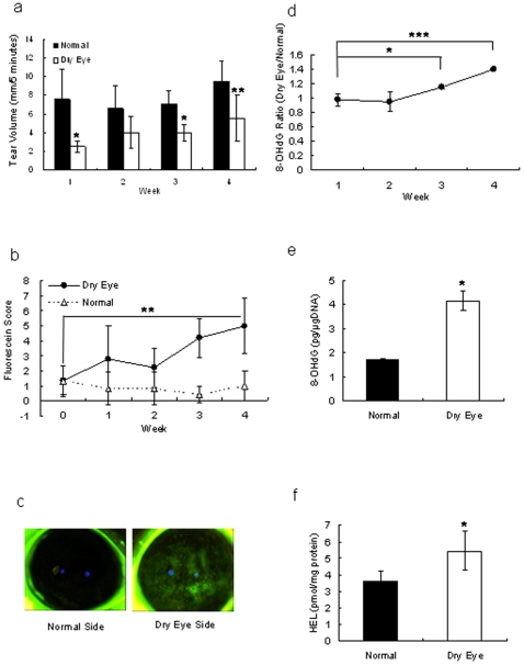
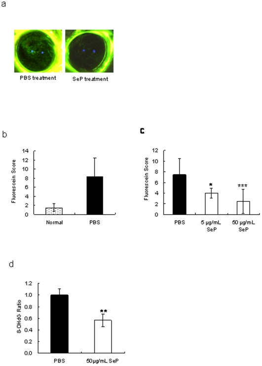
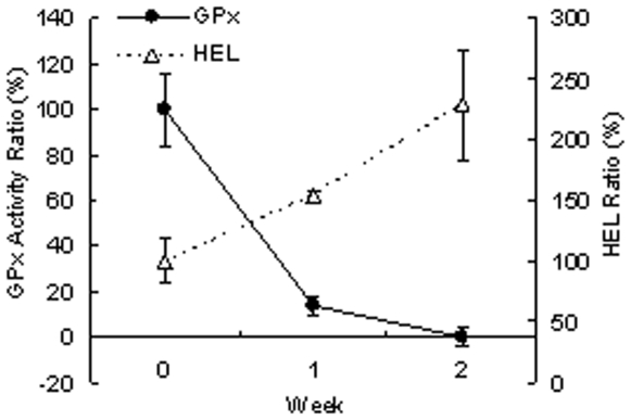
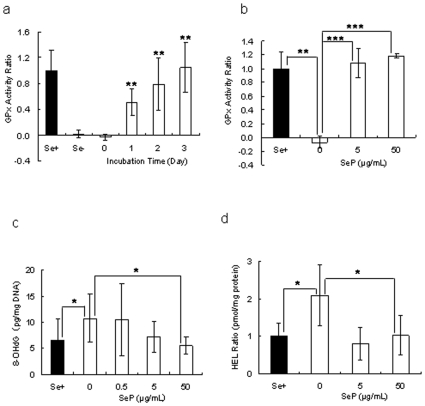
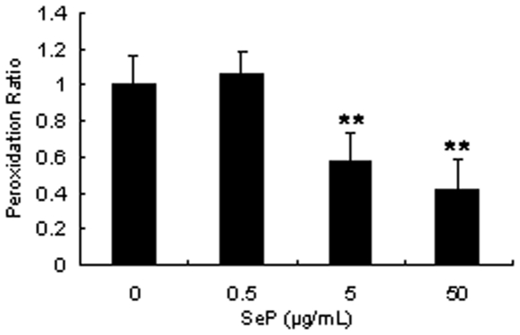
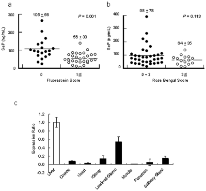
The ocular surface is always attacked by oxidative stress, and cornea epithelial cells are supposed to have their own recovery system against oxidative stress. Therefore we hypothesized that tears supply key molecules for preventing oxidative stress in cornea. The potential target key molecule we focused is selenoprotein P (SeP). SeP is a carrier of selenium, which is an essential trace element for many animals, for oxidative stress metabolism in the organism, and was extremely expressed in lacrimal gland. An experiment was performed with SeP eye drops in a rat dry eye model, prepared by removing the lacrimal glands. The anticipated improvement in corneal dry eye index and the suppression of oxidative stress markers were observed in SeP eye drop group. Furthermore, the concentration of SeP was significantly higher in dry eye patients compared with normal volunteers. Collectively, we concluded that tear SeP is a key molecule to protect the ocular surface cells against environmental oxidative stress.
Conflict of interest statement
Figures
Similar articles
-
Selenium compound protects corneal epithelium against oxidative stress.PLoS One. 2012;7(9):e45612. doi: 10.1371/journal.pone.0045612. Epub 2012 Sep 25. PLoS One. 2012. PMID: 23049824 Free PMC article.
-
Alpha-lipoic acid restores tear production in an animal model of dry eye.Exp Eye Res. 2014 Mar;120:1-9. doi: 10.1016/j.exer.2013.12.014. Epub 2014 Jan 4. Exp Eye Res. 2014. PMID: 24394592
-
Is the main lacrimal gland indispensable? Contributions of the corneal and conjunctival epithelia.Surv Ophthalmol. 2016 Sep-Oct;61(5):616-27. doi: 10.1016/j.survophthal.2016.02.006. Epub 2016 Mar 9. Surv Ophthalmol. 2016. PMID: 26968256 Review.
-
Preservation of tear film integrity and inhibition of corneal injury by dexamethasone in a rabbit model of lacrimal gland inflammation-induced dry eye.J Ocul Pharmacol Ther. 2005 Apr;21(2):139-48. doi: 10.1089/jop.2005.21.139. J Ocul Pharmacol Ther. 2005. PMID: 15857280
-
Autologous Serum and Serum Components.Invest Ophthalmol Vis Sci. 2018 Nov 1;59(14):DES121-DES129. doi: 10.1167/iovs.17-23760. Invest Ophthalmol Vis Sci. 2018. PMID: 30481816 Review.
Cited by
-
Effect of aerobic exercise alone or combined with Mediterranean diet on dry eye in obese hypertensive elderly.Ir J Med Sci. 2023 Dec;192(6):3151-3161. doi: 10.1007/s11845-023-03387-6. Epub 2023 May 9. Ir J Med Sci. 2023. PMID: 37160570 Free PMC article. Clinical Trial.
-
Selenium compound protects corneal epithelium against oxidative stress.PLoS One. 2012;7(9):e45612. doi: 10.1371/journal.pone.0045612. Epub 2012 Sep 25. PLoS One. 2012. PMID: 23049824 Free PMC article.
-
Beneficial effect of daidzin in dry eye rat model through the suppression of inflammation and oxidative stress in the cornea.Saudi J Biol Sci. 2018 May;25(4):832-837. doi: 10.1016/j.sjbs.2016.11.016. Epub 2016 Dec 3. Saudi J Biol Sci. 2018. PMID: 29740252 Free PMC article.
-
Oxidative stress and its downstream signaling in aging eyes.Clin Interv Aging. 2014 Apr 11;9:637-52. doi: 10.2147/CIA.S52662. eCollection 2014. Clin Interv Aging. 2014. PMID: 24748782 Free PMC article. Review.
-
Corneal sensitivity following lacrimal gland excision in the rat.Invest Ophthalmol Vis Sci. 2015 May;56(5):3347-54. doi: 10.1167/iovs.15-16717. Invest Ophthalmol Vis Sci. 2015. PMID: 26024120 Free PMC article.
References
-
- Tsubota K, Higuchi A. Serum application for the treatment of ocular surface disorders. Int Ophthalmol Clin. 2000;40:113–122. - PubMed
-
- Craig JP, Singh I, Tomlinson A, Morgan PB, Efron N. The role of tear physiology in ocular surface temperature. Eye. 2000;14:635–641. - PubMed
-
- Cekic O, Ohji M, Hayashi A, Fang XY, Kusaka S, et al. Effects of humidified and dry air on corneal endothelial cells during vitreal fluid-air exchange. Am J Ophthalmol. 2002;134:75–80. - PubMed
-
- Zuclich JA, Connolly JS. Ocular damage induced by near-ultraviolet laser radiation. Invest Ophthalmol Vis Sci. 1976;15:760–764. - PubMed
-
- Nakamura S, Shibuya M, Nakashima H, Hisamura R, Masuda N, et al. Involvement of oxidative stress on corneal epithelial alterations in a blink-suppressed dry eye. Invest Ophthalmol Vis Sci. 2007;48:1552–1558. - PubMed
Publication types
MeSH terms
Substances
LinkOut - more resources
Full Text Sources
Other Literature Sources
Medical
