. 2010 Mar 26;5(3):e9891.
doi: 10.1371/journal.pone.0009891.

Delayed resolution of acute inflammation in ulcerative colitis is associated with elevated cytokine release downstream of TLR4

Affiliations

Delayed resolution of acute inflammation in ulcerative colitis is associated with elevated cytokine release downstream of TLR4

Farooq Z Rahman et al. PLoS One. .

Abstract

Background: Ulcerative colitis (UC) is widely viewed as a leukocyte-mediated disorder. Although strong evidence implicates an exuberant response to microbial components in its pathogenesis, no intrinsic immune defect has been identified and the underlying pathogenic mechanisms remain obscure.

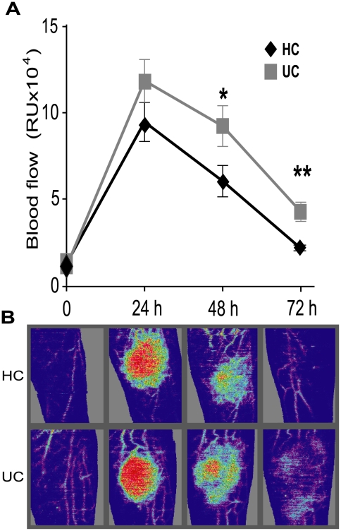
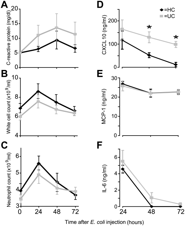
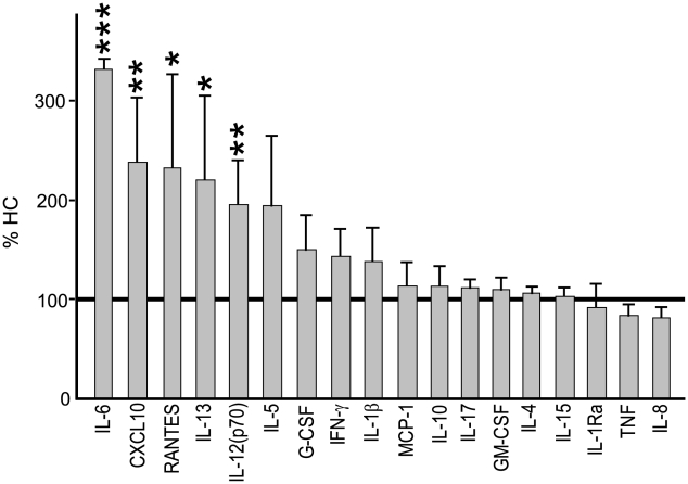
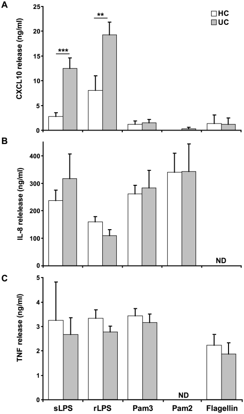
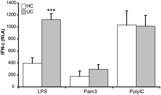
Methodology/principal findings: The acute immune response to bacterial injection was determined in UC patients with quiescent disease and directly compared to healthy control subjects. Monocyte-derived macrophages were used to investigate bacterial recognition mechanisms in vitro. An exuberant and protracted acute inflammatory response to bacteria was evident in patients with UC, which coincides with increased systemic levels of CXCL10. Macrophages stimulated with bacteria and Toll-like receptor (TLR) ligands revealed a specific defect in the TLR4 response in UC. The defect resulted in the over-expression of a number of pro-inflammatory molecules under transcriptional control of the adaptor TIR-domain containing adaptor inducing interferon-beta (TRIF).

Conclusion: These findings highlight a dysregulated innate immune response with over-expression of molecules associated with leukocyte recruitment and activation that may eventuate in the hallmark chronic immune-mediated inflammation of UC.


Conflict of interest statement

Competing Interests: The authors have declared that no competing interests exist.

Figures

Figure 1
Figure 1. Local inflammation was protracted in UC patients after subcutaneous injection of HkEc.
(A) Blood flow at the injection site was elevated and prolonged in patients with UC (n = 21) at 48 (p = 0.04) and 72 h (p = 0.002) compared to HC (n = 15). (B) Laser Doppler images showing elevated and prolonged local blood flow in a UC patient after HkEc inoculation (Red represents high and purple low blood flow).
Figure 2
Figure 2. Systemic CXCL10 levels were elevated in UC patients.
Effects of subcutaneous injection of HkEc on, (A) C-reactive protein (all subjects had baseline CRP levels of <5 mg/dl), (B) white cell count, (C) circulating neutrophil numbers, (D) CXCL10, (E) MCP and (F) -1 IL-6. TNF-α, IFN-γ, IL-8, IL-10, IL-13, IL-17 and MIP-1α were undetectable in serum samples at all time-points. Results are expressed as a mean ± SEM (*p<0.05).
Figure 3
Figure 3. Pro-inflammatory cytokine secretion by UC macrophages (n = 11) in response to HkEc.
Results are expressed as a percentage (mean ± SEM) of that secreted by HC cells (absolute values for HC shown in Table S1). (*p<0.05, **p<0.01 and ***p<0.001).
Figure 4
Figure 4. Macrophages from UC patients demonstrate defective cytokine release downstream of TLR4.
Macrophages from UC (n = 12) and HC (n = 8) were stimulated for 24 h with sLPS (TLR4/CD14), rLPS (TLR4), Pam3CSK4 (TLR2/1), Pam2CSK4 (TLR2/6) and flagellin (TLR5). Secreted levels of (A) CXCL10, (B) IL-8 and (C) TNF-α were measured. Results are expressed as a mean ± SEM (**p<0.01, ***p<0.001) (ND  =  not determined).
Figure 5
Figure 5. TLR4 activation results in an elevation in TRIF signaling and subsequent IFN-β release by macrophage from UC patients.
Macrophages from UC (n = 11) and HC (n = 7) were stimulated for 6 h with rLPS (TLR4), Pam3CSK4 (TLR2/1) or Poly I:C (TLR3) and the levels of IFN-β release in determined. Results are expressed as relative luciferase activity (RLA) (mean ± SEM, ***p<0.001).
Figure 6
Figure 6. TLR-specific gene transcription profiles reveal increased expression of TRIF-responsive genes in macrophages from UC patients after stimulation with HkEc.
Macrophages from UC (n = 8) and HC (n = 8) were stimulated for 4 h with HkEc followed by gene array analysis. (A) Volcano plot showing the alteration in expression levels between UC and HC after HkEc stimulation. Only eight genes (p<0.05 (blue line) and fold change >2) demonstrated significantly different expression levels after stimulation (labeled a–h). (B) Identification of the abnormally expressed genes with the corresponding fold change and p-value.

Similar articles

Cited by

References

    1. Brain O, Travis SP. Therapy of ulcerative colitis: state of the art. Curr Opin Gastroenterol. 2008;24:469–474. - PubMed
    1. Xavier RJ, Podolsky DK. Unravelling the pathogenesis of inflammatory bowel disease. Nature. 2007;448:427–434. - PubMed
    1. Farrell RJ, Peppercorn MA. Ulcerative colitis. Lancet. 2002;359:331–340. - PubMed
    1. Fisher SA, Tremelling M, Anderson CA, Gwilliam R, Bumpstead S, et al. Genetic determinants of ulcerative colitis include the ECM1 locus and five loci implicated in Crohn's disease. Nat Genet. 2008;40:710–712. - PMC - PubMed
    1. Franke A, Balschun T, Karlsen TH, Hedderich J, May S, et al. Replication of signals from recent studies of Crohn's disease identifies previously unknown disease loci for ulcerative colitis. Nat Genet. 2008;40:713–715. - PubMed

Publication types