. 2010 Mar 31:16:556-61.

TGFBI gene mutation analysis in a Chinese pedigree of Reis-Bücklers corneal dystrophy

TGFBI gene mutation analysis in a Chinese pedigree of Reis-Bücklers corneal dystrophy

Ke Ma et al. Mol Vis. .

Abstract

Purpose: To analyze transforming growth factor beta-induced (TGFBI) gene mutations in a Chinese pedigree with Reis-Bücklers dystrophy (RBCD).

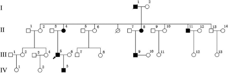
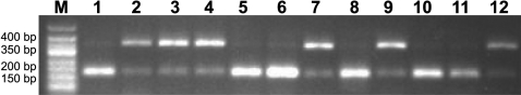
Methods: In a four-generation Chinese family with Reis-Bücklers dystrophy, six members were patients and the rest were unaffected. All members of the family underwent complete ophthalmologic examinations. Exons of TGFBI were amplified by polymerase chain reaction, sequenced, and compared with a reference database. The sequencing results were reconfirmed by polymerase chain reaction-restriction fragment length polymorphism (PCR-RFLP).

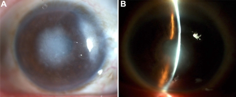
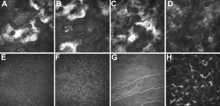
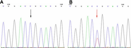
Results: A single heterozygous C>T (R124C) point mutation was found in exon 4 of TGFBI in all six members of the pedigree affected with RBCD, but not in the unaffected members.

Conclusions: Within this pedigree, RBCD segregates with the R124C variance, which is a known mutation for lattice corneal dystrophy type I. Therefore, along with G623D and R124L, the R124C mutation in TGFBI is also found to be responsible for RBCD.


Figures

Figure 1
Figure 1
The pedigree of the family with Reis-Bücklers dystrophy (RBCD). The circle indicates female, the square indicates male, and the filled circle or square indicates the affected individual with RBCD. Arrow signifies the proband and slash through symbol indicates death.
Figure 2
Figure 2
Slit-lamp examination showed an irregular corneal surface and several discrete, gray-white opacities in the subepithelial area and Bowman’s layer. Some of the opacities were associated with marked corneal scarring. No neovascularization and stromal lattice were observed.
Figure 3
Figure 3
In vivo laser scanning confocal microscopy. AD: Patient III:3 36 μm (A), 42 μm (B), 59 μm (C), and 110 μm (D) from the corneal surface. Focal deposition of homogeneous reflective materials with rounded and hyporeflective edges were observed (AC). Amyloid-like deposits that were hyperreflective and with poorly demarcated margins were observed (D). EH: Normal individual: 36 μm (A), 42 μm (B), 57 μm (C), and 113 μm (D) from the corneal surface. Epithelium cells (E, F) nerve fibers (G), and corneal stromal cells (H) were observed.
Figure 4
Figure 4
TGFBI heterozygous mutation in the family. A: Unaffected individuals of the family. B: Patients. The sequence in an affected member shows a heterozygous C>T transversion (indicated by the arrow).
Figure 5
Figure 5
PCR-RFLP. Analysis of R124C mutation by polymerase chain reaction restriction fragment length polymorphism (PCR-RFLP). Lane 1–12: II:2, II:4, II:8, II:11, II:13, III:4, III:5, III:6, III:9, III:13, IV:1, and IV:3 of the pedigree. Lane 2, 3, 4, 7, 9, and 12 are affected individuals, and the rest are unaffected individuals of the pedigree.

Similar articles

Cited by

References

    1. Wittebol-Post D, Pels E. The dystrophy described by Reis and Bücklers. Separate entity or variant of the granular dystrophy? Ophthalmologica. 1989;199:1–9. - PubMed
    1. Kuchle M, Green WR, Volcker HE, Barraquer J. Reevaluation of corneal dystrophies of Bowman's layer and the anterior stroma (Reis-Bucklers and Thiel-Behnke types): a light and electron microscopic study of eight corneas and a review of the literature. Cornea. 1995;14:333–54. - PubMed
    1. Wheeldon CE, De Karolyi BH, Patel DV, Sherwin T, McGhee CN, Vincent AL. A novel phenotype-genotype relationship with a TGFBI exon 14 mutation in a pedigree with a unique corneal dystrophy of Bowman’s layer. Mol Vis. 2008;14:1503–12. - PMC - PubMed
    1. Liu Z, Wang YQ, Gong QH, Xie LX. An R124C mutation in TGFBI caused lattice corneal dystrophy type I with a variable phenotype in three Chinese families. Mol Vis. 2008;14:1234–9. - PMC - PubMed
    1. Munier FL, Frueh BE, Othenin-Girard P, Uffer S, Cousin P, Wang MX, Heon E, Black GC, Blasi MA, Balestrazzi E, Lorenz B, Escoto R, Barraquer R, Hoeltzenbein M, Gloor B, Fossarello M, Singh AD, Arsenijevic Y, Zografos L, Schorderet DF. BIGH3 Mutation Spectrum in Corneal Dystrophies. Invest Ophthalmol Vis Sci. 2002;43:949–54. - PubMed

Substances

LinkOut - more resources