. 2010 Mar 26;5(3):e9909.
doi: 10.1371/journal.pone.0009909.

Isoform-specific regulation and localization of the coxsackie and adenovirus receptor in human airway epithelia

Affiliations

Isoform-specific regulation and localization of the coxsackie and adenovirus receptor in human airway epithelia

Katherine J D A Excoffon et al. PLoS One. .

Abstract

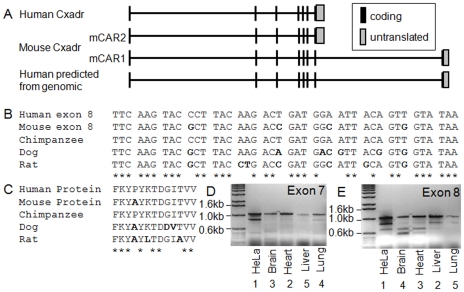
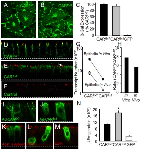
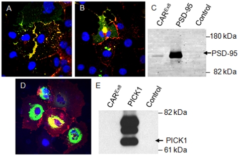
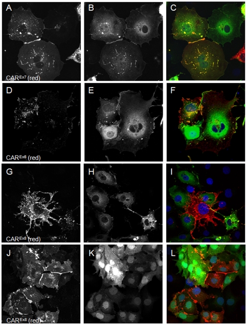
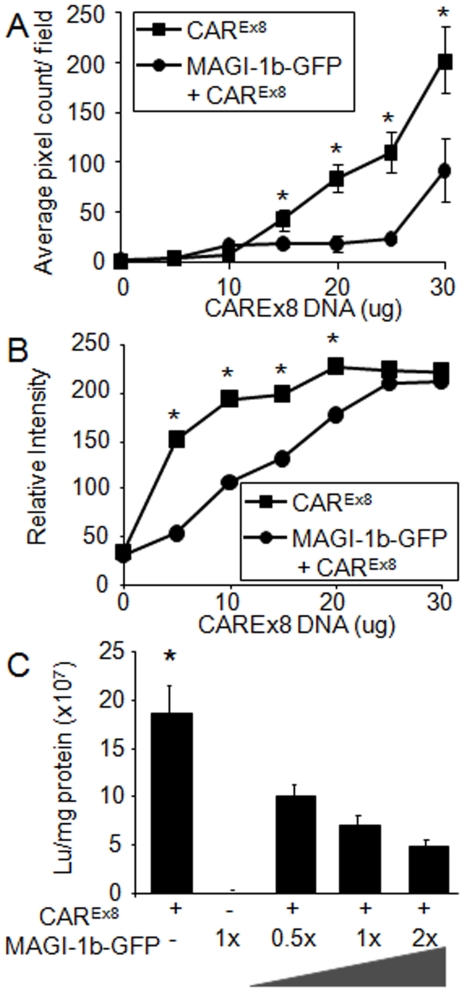
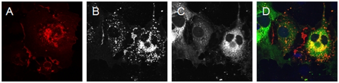
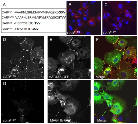
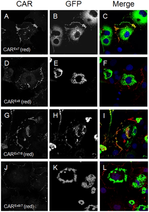
Adenovirus is an important respiratory pathogen. Adenovirus fiber from most serotypes co-opts the Coxsackie-Adenovirus Receptor (CAR) to bind and enter cells. However, CAR is a cell adhesion molecule localized on the basolateral membrane of polarized epithelia. Separation from the lumen of the airways by tight junctions renders airway epithelia resistant to inhaled adenovirus infection. Although a role for CAR in viral spread and egress has been established, the mechanism of initial respiratory infection remains controversial. CAR exists in several protein isoforms including two transmembrane isoforms that differ only at the carboxy-terminus (CAR(Ex7) and CAR(Ex8)). We found low-level expression of the CAR(Ex8) isoform in well-differentiated human airway epithelia. Surprisingly, in contrast to CAR(Ex7), CAR(Ex8) localizes to the apical membrane of epithelia where it augments adenovirus infection. Interestingly, despite sharing a similar class of PDZ-binding domain with CAR(Ex7), CAR(Ex8) differentially interacts with PICK1, PSD-95, and MAGI-1b. MAGI-1b appears to stoichiometrically regulate the degradation of CAR(Ex8) providing a potential mechanism for the apical localization of CAR(Ex8) in airway epithelial. In summary, apical localization of CAR(Ex8) may be responsible for initiation of respiratory adenoviral infections and this localization appears to be regulated by interactions with PDZ-domain containing proteins.


Conflict of interest statement

Competing Interests: The authors have declared that no competing interests exist.

Figures

Figure 1
Figure 1. Human CAR is an 8 exon alternatively spliced protein.
Panel A shows a schematic diagram of the published and predicted human and mouse exon arrangement. Panel B shows the alignment of the mouse CAR exon 8 with the predicted exon 8 from other species. Panel C and D show representative RT-PCR for human CAR Exon 8 or 7, respectively, in cells (HeLa) or tissues.
Figure 2
Figure 2. Human CAREx8 localization and adenovirus-mediated gene transfer is similar to hCAREx7 in cell monolayers but distinct in polarized cells.
COS-7 cells transfected with hCAREx7 (A) or hCAREx8 (B) show a similar distribution. Panel C shows that CHO cells transfected with hCAREx7 or hCAREx8 mediate similar adenovirus gene transfer. Immunocytochemistry for endogenous hCAREx7 (D) or hCAREx8 (E) (green) reveals distinct localization in polarized primary human airway epithelia greater than 2 weeks of age. hCAREx7 localizes to the basolateral membrane and shows co-localization with the basolateral portion of ZO-1 (red, D, E, F). hCAREx8 localizes diffusely in the upper region of the cytoplasm with some apical staining (see arrowhead). Panel F shows ZO-1 (red) and a lack of staining with control rabbit pre-immune serum (green). Panel G shows the abundance of CAREx7 or CAREx8 transcripts in primary cultures (in vitro) or from lung tissue (in vivo). Panel H shows that the relative enrichment of CAREx7 to CAREx8 transcripts is similar in vitro and in vivo. Localization of hCAREx7 (I) or hCAREx8 (J, K, L, M) after over-expression in primary cultures and co-stained (red) for ZO-1 (I, J), acetylated α-tubulin (K), CD55/decay accelerating factor (DAF, L), or ezrin (M). Panel N shows that expression of exogenous hCAREx8 in polarized human airway epithelia mediates two-fold greater adenovirus gene transfer than hCAREx7 in comparison to control GFP transduced cells. *p<0.01. Confocal microscopy (60x oil immersion).
Figure 3
Figure 3. hCAREx8 co-localizes and interacts with PSD-95 but not PICK1.
Panel A shows co-localization (yellow) of hCAREx8 (red) and PSD-95-GFP (green). In contrast, in panel B, hCAREx8-PDZ does not co-localize at the junctions of cells. hCAREx8-PDZ localizes to the junctions between cells whereas PSD-95-GFP fluorescence remains diffuse. Panel C shows immunoprecipitation of PSD-95-GFP with the hCAR specific extracellular domain monoclonal antibody RmcB, GFP antibody, but not a control antibody (MopC). Panels D and E shows the lack of co-localization and immunoprecipitation between hCAREx8 (junctional) and PICK1-GFP (perinuclear). Confocal microscopy (60x oil immersion).
Figure 4
Figure 4. Co-expression of hCAREx8 and MAGI-1b-GFP results in the loss of hCAREx8.
In contrast to the co-localization of hCAREx7 (A, red) and MAGI-1b-GFP (B, green) as shown in panel C (yellow), co-expression of hCAREx8 (D, G, red) and MAGI-1b-GFP (E, H, green) results in decreased levels of hCAREx8 (F) unless MAGI-1b-GFP is absent from the cell (I). Co-expression of hCAREx8 (J, red) with GFP (K, green) results in abundant hCAREx8 expression at the junctions of the cells and diffuse GFP expression (L). Confocal microscopy (60x oil immersion).
Figure 5
Figure 5. Co-expression of hCAREx8 with MAGI-1b-GFP results in less immunofluorescence, protein, and adenovirus-mediated gene transfer.
COS-7 cells were transfected with 0 to 30 µg of hCAREx8 +/− 15 µg MAGI-1b-GFP and evaluated for hCAREx8 specific immunofluorescence (A) or CAREx8 specific protein by Western blot (B). In the presence of MAGI-1b-GFP, the hCAREx8 expression curve is shifted to the right suggesting a loss of hCAREx8 protein. Panel C shows CHO cells transfected with varying amounts of hCAREx8 and MAGI-1b-GFP, and evaluated for Ad-β-galactosidase gene transfer. Co-expression of MAGI-1b-GFP resulted in a decrease of adenovirus-mediated gene transfer in a dose response relationship. *p<0.03.
Figure 6
Figure 6. MAGI-1b-GFP-mediated loss of hCAREx8 requires the PDZ-binding domain (ITVV) of hCAREx8.
COS-7 cells were transfected with hCAREx8-PDZ (A, red) or co-transfected with hCAREx8-PDZ (B, red) and MAGI-1b-GFP (C, green). Panel D shows the lack of co-localization of hCAREx8-PDZ (junctions) and MAGI-1b-GFP (cytoplasmic/diffuse). Confocal microscopy (60x oil immersion).
Figure 7
Figure 7. The result of the MAGI-1b-GFP interaction with hCAR requires both the PDZ binding domain and the upstream isoform specific amino acids.
The PDZ binding domain of hCAREx7 and hCAREx8 were swapped by PCR as shown in panel A. Both constructs contained identical upstream sequences. The localization was determined in transfected COS-7 cells either alone, hCAREx7/8 (B), hCAREx8/7 (C) or upon co-transfection with MAGI-1b-GFP (D-I). Panels D and E show junctional expression of hCAREx7/8 and MAGI-1b-GFP, respectively, and co-localization in panel F. Panel G shows minor expression of hCAREx8/7 in the presence of robust MAGI-1b-GFP expression in panel H. Some co-localization is observed in panel I. Confocal microscopy (60x oil immersion).
Figure 8
Figure 8. The interaction between the hCAR PDZ binding domain and PICK1 depends on both the PDZ binding domain and the upstream isoform specific amino acids.
The PDZ binding domain of hCAREx7 and hCAREx8 were swapped by PCR as shown in Figure 7, panel A. The localization was determined in transfected COS-7 cells. hCAREx7 (A) transfected with PICK1-GFP (B) results in co-localization at the junctions (C, yellow). hCAREx8 (D) transfected with PICK1-GFP (E) results in no co-localization at the junctions (F). hCAREx7/8 (G) transfected with PICK1-GFP (H) results some co-localization at the junctions (I, yellow). hCAREx8/7 (J) transfected with PICK1-GFP (K) results in no co-localization at the junctions (L). Confocal microscopy (60x oil immersion).

References

    1. Bergelson JM, Cunningham JA, Droguett G, Kurt-Jones EA, Krithivas A, et al. Isolation of a common receptor for coxsackie B viruses and adenoviruses 2 and 5. Science. 1997;275:1320–1323. - PubMed
    1. Tomko RP, Xu R, Philipson L. HCAR and MCAR: the human and mouse cellular receptors for subgroup C adenoviruses and group B coxsackieviruses. Proc Natl Acad Sci U S A. 1997;94:3352–3356. - PMC - PubMed
    1. Honda T, Saitoh H, Masuko M, Katagiri-Abe T, Tominaga K, et al. The coxsackievirus-adenovirus receptor protein as a cell adhesion molecule in the developing mouse brain. Brain Res Mol Brain Res. 2000;77:19–28. - PubMed
    1. Walters R, Freimuth P, Moninger T, Ganske I, Zabner J, et al. Adenovirus Fiber Disrupts CAR-Mediated Intercellular Adhesion Allowing Virus Escape. Cell. 2002;110:789–799. - PubMed
    1. Cohen CJ, Shieh JT, Pickles RJ, Okegawa T, Hsieh JT, et al. The coxsackievirus and adenovirus receptor is a transmembrane component of the tight junction. Proc Natl Acad Sci U S A. 2001;98:15191–15196. - PMC - PubMed

Publication types

MeSH terms

LinkOut - more resources