

(-)-Epigallocatechin-3-gallate inhibits Met signaling, proliferation, and invasiveness in human colon cancer cells
- PMID: 20361925
- PMCID: PMC2916072
- DOI: 10.1016/j.abb.2010.03.017
(-)-Epigallocatechin-3-gallate inhibits Met signaling, proliferation, and invasiveness in human colon cancer cells
Abstract
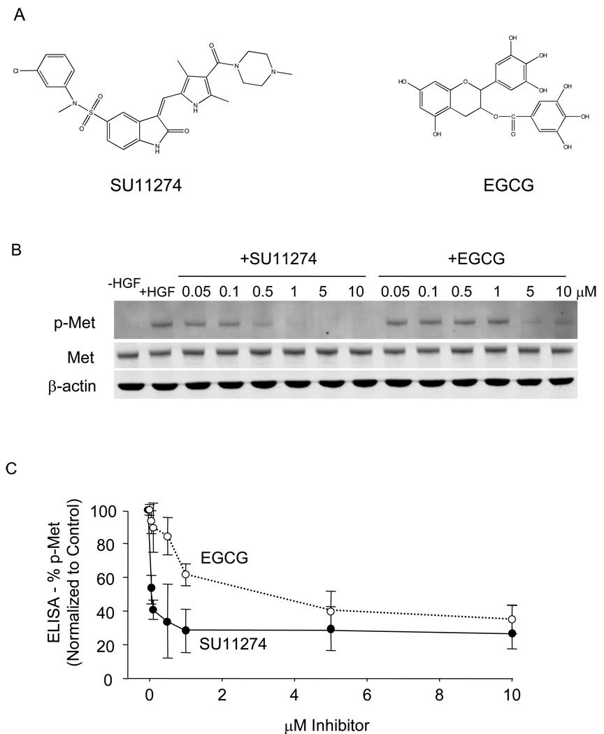
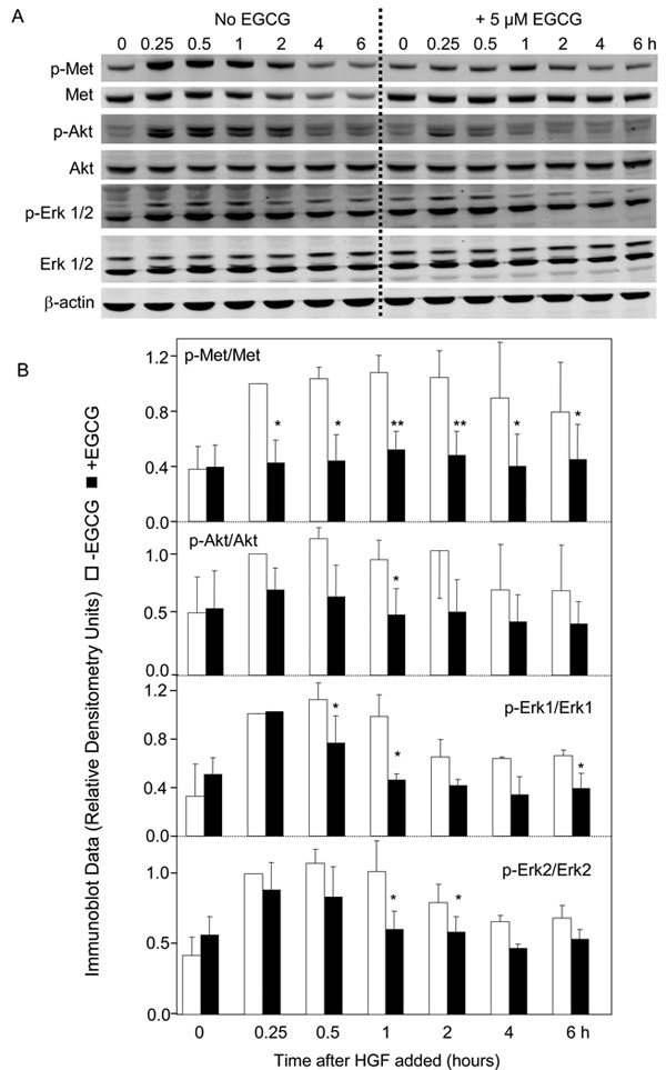
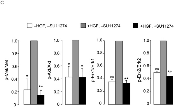
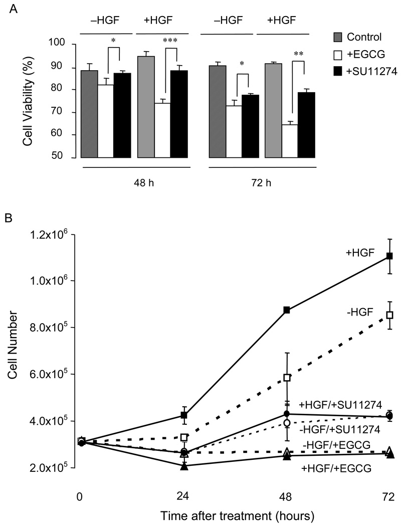
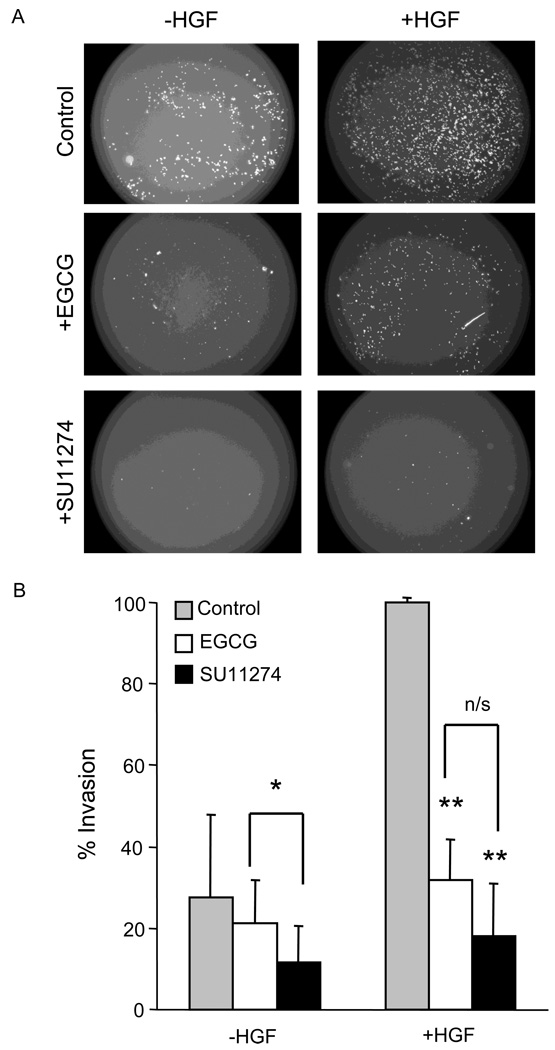
The Met receptor tyrosine kinase is deregulated in a variety of cancers and is correlated with advanced stage and poor prognosis. Thus, Met has been identified as an attractive candidate for targeted therapy. We compared the tea polyphenol (-)-epigallocatechin-3-gallate (EGCG) and a specific Met inhibitor, SU11274, as suppressing agents of Met signaling in HCT116 human colon cancer cells. Treatment with hepatocyte growth factor increased phospho-Met levels, and this was inhibited in a concentration-dependent manner by EGCG and SU11274 (IC(50) 3.0 vs. 0.05muM, respectively). Downstream activation of Erk and Akt signaling pathways also was suppressed. Both compounds at a concentration of 5muM lowered cell viability and proliferation, with EGCG being more effective than SU11274, and the invasion of colon cancer cells in Matrigel assays was strongly inhibited. These findings are discussed in the context of the pleiotropic effects of tea catechins, their tissue metabolite levels, and the potential to inhibit colon cancer metastasis and invasion.
2010 Elsevier Inc. All rights reserved.
Figures
Similar articles
-
The green tea polyphenol EGCG potentiates the antiproliferative activity of c-Met and epidermal growth factor receptor inhibitors in non-small cell lung cancer cells.Clin Cancer Res. 2009 Aug 1;15(15):4885-94. doi: 10.1158/1078-0432.CCR-09-0109. Epub 2009 Jul 28. Clin Cancer Res. 2009. PMID: 19638461 Free PMC article.
-
The green tea catechins, (-)-Epigallocatechin-3-gallate (EGCG) and (-)-Epicatechin-3-gallate (ECG), inhibit HGF/Met signaling in immortalized and tumorigenic breast epithelial cells.Oncogene. 2006 Mar 23;25(13):1922-30. doi: 10.1038/sj.onc.1209227. Oncogene. 2006. PMID: 16449979
-
Green tea (-)-epigallocatechin-3-gallate inhibits HGF-induced progression in oral cavity cancer through suppression of HGF/c-Met.J Nutr Biochem. 2011 Nov;22(11):1074-83. doi: 10.1016/j.jnutbio.2010.09.005. Epub 2011 Feb 2. J Nutr Biochem. 2011. PMID: 21292466
-
Effect of c-Met inhibitor SU11274 on human colon cancer cell growth.Chin Med J (Engl). 2013 Jul;126(14):2705-9. Chin Med J (Engl). 2013. PMID: 23876900
-
Suppression of Met activation in human colon cancer cells treated with (-)-epigallocatechin-3-gallate: minor role of hydrogen peroxide.Biochem Biophys Res Commun. 2009 Nov 20;389(3):527-30. doi: 10.1016/j.bbrc.2009.09.019. Epub 2009 Sep 8. Biochem Biophys Res Commun. 2009. PMID: 19744467 Free PMC article.
Cited by
-
Molecular Targets of Natural Compounds with Anti-Cancer Properties.Int J Mol Sci. 2021 Dec 20;22(24):13659. doi: 10.3390/ijms222413659. Int J Mol Sci. 2021. PMID: 34948455 Free PMC article. Review.
-
Advances in the Antagonism of Epigallocatechin-3-gallate in the Treatment of Digestive Tract Tumors.Molecules. 2019 May 3;24(9):1726. doi: 10.3390/molecules24091726. Molecules. 2019. PMID: 31058847 Free PMC article. Review.
-
Tea catechins as inhibitors of receptor tyrosine kinases: mechanistic insights and human relevance.Pharmacol Res. 2010 Dec;62(6):457-64. doi: 10.1016/j.phrs.2010.07.010. Epub 2010 Aug 4. Pharmacol Res. 2010. PMID: 20691268 Free PMC article. Review.
-
Chromatin accessibility underlies synthetic lethality of SWI/SNF subunits in ARID1A-mutant cancers.Elife. 2017 Oct 2;6:e30506. doi: 10.7554/eLife.30506. Elife. 2017. PMID: 28967863 Free PMC article.
-
Involvement of Met receptor pathway in aggressive behavior of colorectal cancer cells induced by parathyroid hormone-related peptide.World J Gastroenterol. 2022 Jul 14;28(26):3177-3200. doi: 10.3748/wjg.v28.i26.3177. World J Gastroenterol. 2022. PMID: 36051345 Free PMC article.
References
-
- Naran S, Zhang X, Hughes SJ. Inhibition of HGF/MET as therapy for malignancy. Expert Opin. Ther. Targets. 2009;13:569–581. - PubMed
-
- Birchmeier C, Birchmeier W, Gherardi E, Vande Woude GF. Met, metastasis, motility and more. Nat. Rev. Mol. Cell Biol. 2003;4:915–925. - PubMed
-
- Christensen JG, Burrows J, Salgia R. c-Met as a target for human cancer and characterization of inhibitors for therapeutic intervention. Cancer Lett. 2005;225:1–26. - PubMed
-
- Fujita S, Sugano K. Expression of c-met proto-oncogene in primary colorectal cancer and liver metastases. Jpn. J. Clin. Oncol. 1997;27:378–383. - PubMed
-
- Jeffers M, Rong S, Woude GF. Hepatocyte growth factor/scatter factor-Met signaling in tumorigenicity and invasion/metastasis. J. Mol. Med. 1996;74:505–513. - PubMed
Publication types
MeSH terms
Substances
Grants and funding
LinkOut - more resources
Full Text Sources
Miscellaneous
