. 2010 Apr 9;86(4):639-49.
doi: 10.1016/j.ajhg.2010.03.002. Epub 2010 Apr 1.

Severe X-linked mitochondrial encephalomyopathy associated with a mutation in apoptosis-inducing factor

Affiliations

Severe X-linked mitochondrial encephalomyopathy associated with a mutation in apoptosis-inducing factor

Daniele Ghezzi et al. Am J Hum Genet. .

Abstract

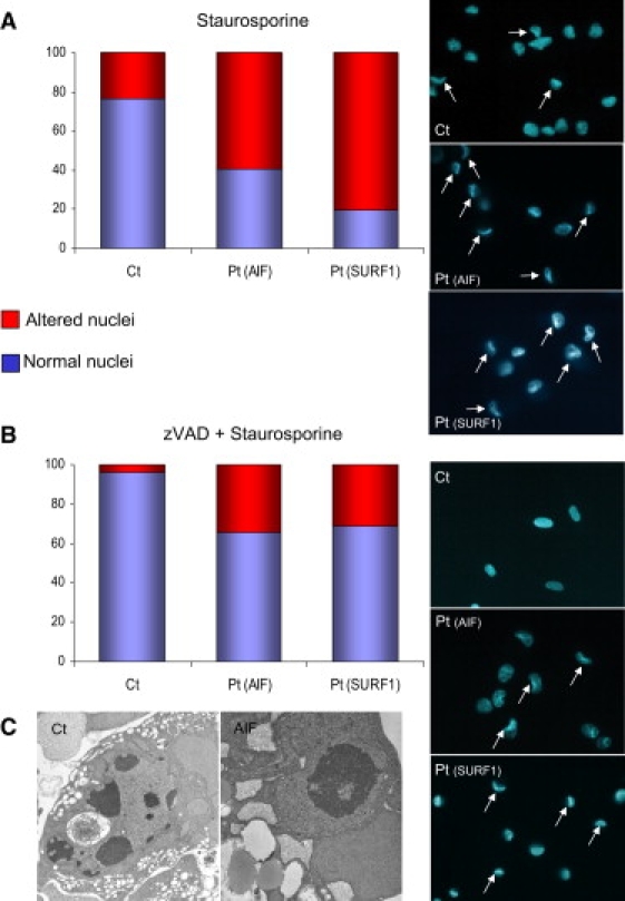
We investigated two male infant patients who were given a diagnosis of progressive mitochondrial encephalomyopathy on the basis of clinical, biochemical, and morphological features. These patients were born from monozygotic twin sisters and unrelated fathers, suggesting an X-linked trait. Fibroblasts from both showed reduction of respiratory chain (RC) cIII and cIV, but not of cI activities. We found a disease-segregating mutation in the X-linked AIFM1 gene, encoding the Apoptosis-Inducing Factor (AIF) mitochondrion-associated 1 precursor that deletes arginine 201 (R201 del). Under normal conditions, mature AIF is a FAD-dependent NADH oxidase of unknown function and is targeted to the mitochondrial intermembrane space (this form is called AIF(mit)). Upon apoptogenic stimuli, a soluble form (AIF(sol)) is released by proteolytic cleavage and migrates to the nucleus, where it induces "parthanatos," i.e., caspase-independent fragmentation of chromosomal DNA. In vitro, the AIF(R201 del) mutation decreases stability of both AIF(mit) and AIF(sol) and increases the AIF(sol) DNA binding affinity, a prerequisite for nuclear apoptosis. In AIF(R201 del) fibroblasts, staurosporine-induced parthanatos was markedly increased, whereas re-expression of AIF(wt) induced recovery of RC activities. Numerous TUNEL-positive, caspase 3-negative nuclei were visualized in patient #1's muscle, again indicating markedly increased parthanatos in the AIF(R201 del) critical tissues. We conclude that AIF(R201 del) is an unstable mutant variant associated with increased parthanatos-linked cell death. Our data suggest a role for AIF in RC integrity and mtDNA maintenance, at least in some tissues. Interestingly, riboflavin supplementation was associated with prolonged improvement of patient #1's neurological conditions, as well as correction of RC defects in mutant fibroblasts, suggesting that stabilization of the FAD binding in AIF(mit) is beneficial.


Figures

Figure 1
Figure 1
Brain Imaging and Skeletal Muscle Morphological and Histochemical Analysis (A and B) Magnetic resonance imaging (MRI) T2-weighted (A) and T1-weighted (B) sequences on a transverse section of supratentorial brain of patient #1. (C) MRI T2-weighted sequence on a transverse section of supratentorial brain of patient #2 at 11 months of age. (D) MRI T1-weighted sequence on a coronal section of supratentorial brain of patient #2 at 11 months of age. (E–H) Patient #2 muscle biopsy showing an increase of connective tissue, severe variability in fiber size, and necrotic and regenerating fibers at Hematoxylin and Eosin (H&E) (E), numerous ragged-red fibers under the modified Gomori trichrome staining (F), increased SDH (succinate dehydrogenase) histochemical staining (G), and diffuse reduction of COX histochemical reaction (H).
Figure 2
Figure 2
Pedigree and AIF Features (A) Pedigree of the family. Filled symbols indicate affected individuals (patient #1, Pt#1 and patient #2, Pt#2, also indicated by arrows). WT and ΔR201 indicate the presence of wild-type AIF sequence and the presence of the R201 deletion, respectively. (B) Sequence analysis of exon 5 of AIFM1 showing the c.601–603 deletion in patient #1. A control sequence is shown for comparison. (C) Schematic primary structure of the AIF protein. MTS indicates the mitochondrial targeting sequence; TM indicates a putative transmembrane domain. MPP: Mitochondrial Processing Peptidase. The precursor, mitochondrial (AIFmit), and soluble (AIFsol) forms of the AIF protein are shown starting from the N-terminal aminoacid; the corresponding molecular weights are also indicated.
Figure 3
Figure 3
Structural and In Vitro Analyses of Recombinant AIFwt and AIF201 del (A) Superimposed X-ray structures of oxidized (NAD-less) human and murine AIFs (PDB codes: 1M6I and 1DV416,17) show that the R201 (R200 in mouse) assists in the folding of two peptide stretches, near the FAD-binding pocket. The R201 residue, contained in the first stretch (in pink), establishes a link with the second stretch (in brown) by forming a salt bridge with the E531 residue. (B) In the reduced NAD-bound dimer of murine AIF (PDB code 3GD418), the active form of the flavoprotein, R200 (R201 in humans), establishes a hydrogen bond with F204, helping the functionally important β-hairpin, including the W195 residue (shown in pink), to get properly oriented. F204 and W195 correspond to human F205 and W196, respectively. (C) Computer modeling predicts that deletion of R201 in reduced human AIF can disrupt the H bond with F205 and thus shorten and distort the 191–203 β-hairpin (rendered in blue for the wild-type and in pink for the mutant). (D) Anaerobic titration of 15 μM AIFmitR201 del with NADH (in 50 mM phosphate [pH 7.5]). The inset shows that an equimolar amount of NADH is required to fully reduce FAD. (E) Oxidation of the NADH-reduced wild-type (wt) and R201 del mutants (mt) of AIFmit and AIFsol. A 15 μM protein solution in 50 mM phosphate (pH 7.5) was aerobically reduced with a 6-fold excess of NADH, and the FAD oxidation by air oxygen was monitored over time at 452 nm. (F) Far-UV circular dichroism spectra of 3 μM AIFmitR201 del were recorded in 10 mM sodium phosphate (pH 7.5) in the absence or presence of a 20-fold excess of NAD(P)H. The R201 del mutation has no significant effect on the redox-dependent changes in the secondary structure of AIF. (G) The R201 del mutation promotes the AIFsol-DNA interaction. Equal amounts of wild-type (wt) and mutant (mt) AIFsol were incubated for 15 min with 250 ng of a 100 bp DNA ladder in the absence or presence of a 20-fold excess of NAD(P)H; separation on a 2% agarose gel and visualization with ethidium bromide followed. The R201 deletion was introduced into the AIFmit and AIFsol expression plasmids by site-directed mutagenesis with the Stratagene QuikChange kit. The mutant proteins were expressed and purified according to the procedure developed for wild-type AIF.
Figure 4
Figure 4
Characterization of Patients' Fibroblasts before and after Galactose Treatment (A) Protein-blot analysis of patient #1(Pt) and control (Ct) skin cultured fibroblasts, immunodetected with an antibody against AIF (Chemicon). Antibodies against subunit A of complex II (SDHA, Invitrogen) and actin (Sigma) were used as loading controls. (B) Representative images of mitochondrial morphology, showing the filamentous (upper panel) or the fragmented (lower panel) mitochondrial network of fibroblasts grown in DMEM-glucose or DMEM-galactose. Cells were stained with 100 nM Mitotracker-Red CMX-Ros (Invitrogen) for 1/2 hr. The scale bar represents 10 μm. (C) Quantification of fibroblasts with a normal or fragmented mitochondrial network in controls (Ct) and patients (Pt) after incubation in D-MEM +5 mM galactose for 48 hr. (D) MTT cell-viability assay. Control (Ct, blue lines) and patients' (Pt#1, red lines, and Pt#2, brown lines) fibroblasts were grown in DMEM + glucose (solid lines) or DMEM + galactose (dotted lines) for the times indicated. Absorbance (Abs) is proportional to the number of viable cells.
Figure 5
Figure 5
Staurosporine-Induced Cell Death (A) Quantification of altered nuclei in control fibroblasts (Ct), in fibroblasts from patient #1, Pt(AIF) and from a patient with SURF1 mutations, Pt(SURF1) after staurosporine treatment (1 mM for 2 hr). The three panels on the right show representative fields of Hoechst-stained nuclei. White arrows indicate cells with collapsed nuclei. (B) Samples were the same as in (A), but the treatment consisted of zVAD (100 μM for 1/2 hr) + staurosporine (1 mM for 2 hr). As in (A), the three panels on the right show representative fields of Hoechst-stained nuclei. White arrows indicate cells with collapsed nuclei. (C) Electron microscopy (EM) image of staurosporine-treated fibroblasts. Left panel: a control fibroblast shows apoptotic bodies in the nucleus, typical of caspase-dependent apoptosis; right panel: a fibroblast from patient #1 shows chromatin condensation of the nucleus, typical of parthanatos.
Figure 6
Figure 6
TUNEL, Caspase 3 Immunostaining, and EM in Skeletal Muscle (A, B, and D–F) TUNEL + DAPI staining of skeletal muscle nuclei in skeletal muscle from patient #2 and three disease controls (DMD: from a patient with Duchenne muscular dystrophy; mtDNA del: from a patient with a large heteroplasmic deletion of mtDNA; TK2: from a patient who is compound heterozygote for two pathogenic mutations in the TK2 gene). TUNEL-positive nuclei are in green, whereas TUNEL-negative nuclei are in blue, corresponding to DAPI staining. Numerous TUNEL-positive are present in patient #2; two TUNEL-positive green nuclei are present in the DMD muscle; no TUNEL-positive nuclei are detected in mtDNA del or TK muscle specimens. (C) Anti-Caspase 3 immunostaining of muscle from patient #2 revealed by the diamino-benzidine (DAB) method. No Cas-3-positive fibers were detected. In the insets of (B), (D), and (F), we show the same anti-Cas 3 immunostaining carried out in the corresponding muscle specimens. Only the DMD muscle shows a few Cas-3-positive, brown fibers. (E) EM image of a nucleus with chromatin condensation in muscle tissue from patient #2.

References

    1. Schapira A.H. Mitochondrial disease. Lancet. 2006;368:70–82. - PubMed
    1. Smeitink J.A., Zeviani M., Turnbull D.M., Jacobs H.T. Mitochondrial medicine: A metabolic perspective on the pathology of oxidative phosphorylation disorders. Cell Metab. 2006;3:9–13. - PubMed
    1. Regev-Rudzki N., Pines O. Eclipsed distribution: A phenomenon of dual targeting of protein and its significance. Bioessays. 2007;29:772–782. - PubMed
    1. McFarland R., Turnbull D.M. Batteries not included: Diagnosis and management of mitochondrial disease. J. Intern. Med. 2009;265:210–228. - PubMed
    1. Modjtahedi N., Giordanetto F., Madeo F., Kroemer G. Apoptosis-inducing factor: Vital and lethal. Trends Cell Biol. 2006;16:264–272. - PubMed

Publication types

MeSH terms