. 2010 Jun 20;402(1):61-71.
doi: 10.1016/j.virol.2010.03.013. Epub 2010 Apr 1.

An integrated protein localization and interaction map for Potato yellow dwarf virus, type species of the genus Nucleorhabdovirus

Affiliations

An integrated protein localization and interaction map for Potato yellow dwarf virus, type species of the genus Nucleorhabdovirus

Anindya Bandyopadhyay et al. Virology. .

Abstract

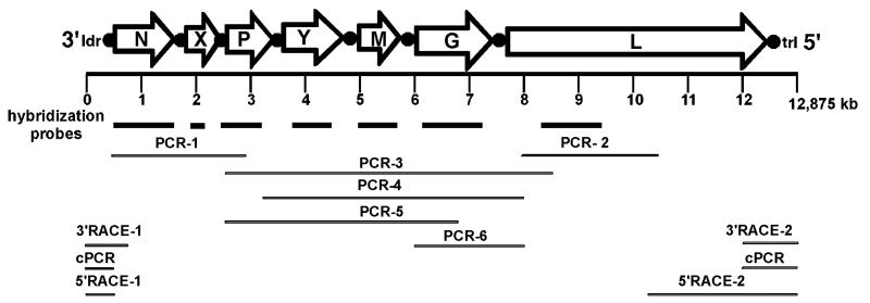
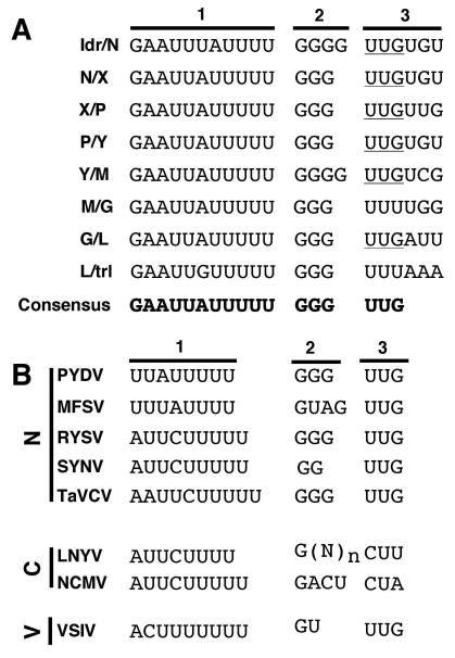
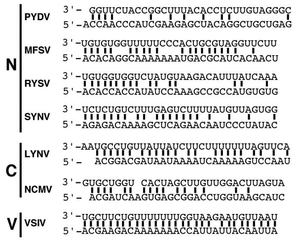
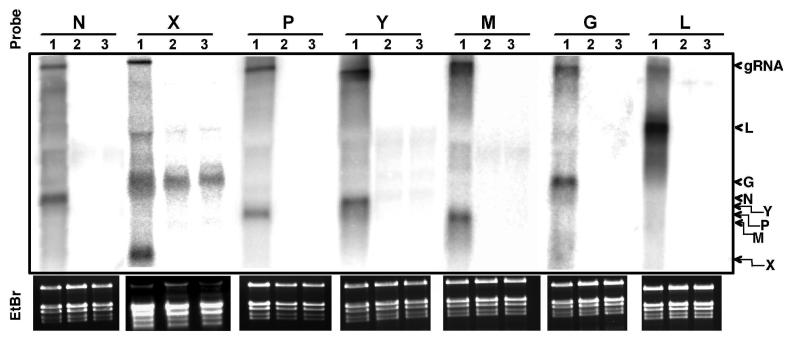
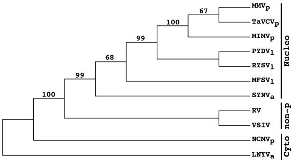
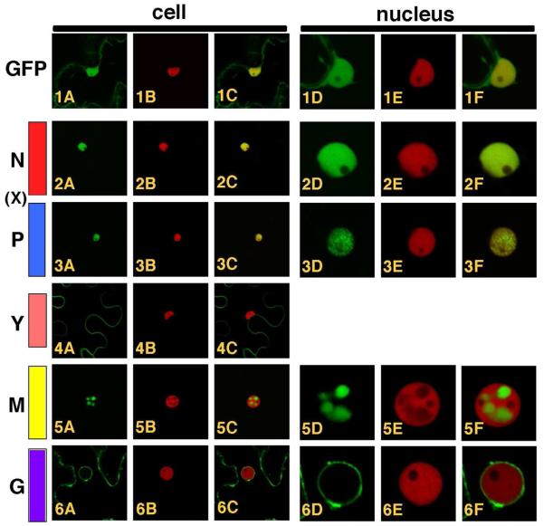
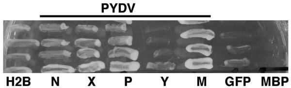
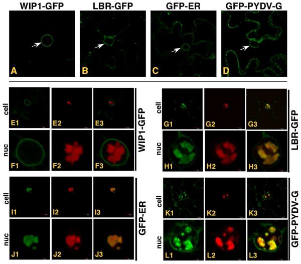
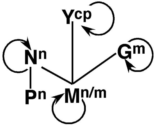
The genome of Potato yellow dwarf virus (PYDV; Nucleorhabdovirus type species) was determined to be 12,875 nucleotides (nt). The antigenome is organized into seven open reading frames (ORFs) ordered 3'-N-X-P-Y-M-G-L-5', which likely encode the nucleocapsid, phospho, movement, matrix, glyco and RNA-dependent RNA polymerase proteins, respectively, except for X, which is of unknown function. The ORFs are flanked by a 3' leader RNA of 149 nt and a 5' trailer RNA of 97 nt, and are separated by conserved intergenic junctions. Phylogenetic analyses indicated that PYDV is closely related to other leafhopper-transmitted rhabdoviruses. Functional protein assays were used to determine the subcellular localization of PYDV proteins. Surprisingly, the M protein was able to induce the intranuclear accumulation of the inner nuclear membrane in the absence of any other viral protein. Finally, bimolecular fluorescence complementation was used to generate the most comprehensive protein interaction map for a plant-adapted rhabdovirus to date.


Figures

Fig. 1
Fig. 1
Organization of the PYDV genome. The 12,875 nt genome encodes seven open reading frames (ORFs; open arrows) that are separated by conserved gene junctions (black circles) and flanked by short leader (ldr) and trailer (trl) sequences, respectively. Probes for each ORF used for nucleic acid hybridization are represented by bold lines. The complete genome sequence was assembled from overlapping fragments generated by standard PCR (PCR), circular PCR (cPCR) or RACE (3′RACE or 5′RACE). Each genome fragment was sequenced at least twice.
Fig. 2
Fig. 2
(A) Sequence of each intergenic junction (IGJ) in the PYDV genomic RNA (drawn here in genomic orientation). The IGJs are divided into three sections to denote the (1) poly-adenylation signal, (2) intergenic spacer and (3) transcription start site. The consensus IGJ is provided at the bottom. (B) Consensus IGJ comparisons from rhabdoviruses in the Nucleorhabdovirus (N), Cytorhabdovirus (C) or Vesiculovirus (V) genera. Abbreviations: (N)n, variable number of nucleotides.
Fig. 3
Fig. 3
Complementary nucleotides (bold lines) in leader (3′) and trailer (5′) terminal sequences of selected rhabdoviruses in the Nucleorhabdovirus (N), Cytorhabdovirus (C) or Vesiculovirus (V) genera.
Fig. 4
Fig. 4
Detection of PYDV transcripts in infected N. benthamiana. Northern gel-blot hybridizations were conducted with ORF-specific probes shown in Fig. 1. 32P-labeled cDNA probes were hybridized to total RNA extracted from PYDV- (lane 1), SYNV-infected (lane 2) or mock-inoculated (lane 3) plants. The relative positions of the PYDV genomic RNA (gRNA) and N, X, P, Y, M, G, and L transcripts are indicated on the right side of this Figure. Ethidium-bromide (EtBr) stained gels at bottom indicate RNA quality and loading for each hybridization. The strong cross-hybridization of the X gene probe with ribosomal RNA
Fig. 5
Fig. 5
Phylogeny of plant rhabdoviruses inferred from L protein sequences. Representative rhabdoviruses infecting a variety of hosts were used, including viruses that do not infect plants (non-p) as well as plant-adapted viruses in the Nucleorhabdovirus (Nucleo) and Cytorhabdovirus (Cyto) genera. Bootstrap values greater than 50% are shown at nodes in the tree. Vectors for the plant-adapted viruses are shown as subscripts, which are aphid (a), leafhopper (l), and planthopper (p). Virus names and Genbank accession numbers are listed in the Materials and
Fig. 6
Fig. 6
Confocal micrographs of PYDV protein fusions expressed by agroinfiltration in leaf epidermal cells of transgenic N. benthamiana plants expressing RFP fused to histone 2B, a nuclear marker. Whole cell or nuclear views of fluorescence of GFP, RFP and overlaid images are provided. 1A-F. GFP. 2A-F. PYDV-N. 3A-F. PYDV-P. 4A-C. PYDV-Y. 5A-F. PYDV-M. 6A-F. PYDV-G. The X protein, shown in its relative genomic location, could not be expressed as a GFP fusion and therefore was omitted here.
Fig. 7
Fig. 7
Yeast-based assay for identification of proteins containing a functional NLS. Positive- (H2B) and negative-control (GFP, MBP) proteins or PYDV proteins (N, X P, Y, and M) were expressed from pNIA-DEST in yeast strain L40. Only those proteins containing a functional NLS (H2B, N, X, P and M) were able to facilitate yeast growth on media lacking histidine.
Fig. 8
Fig. 8
Single plane confocal micrographs of protein fusions of cells in which TagRFP-PYDV-M was co-expressed with various membrane markers fused to GFP by agroinfiltration in leaf epidermal cells of transgenic N. benthamiana plants. White arrows indicate the accumulation of markers on nuclear membranes in the absence of TagRFP-PYDV-M; A. WIP1-GFP (outer nuclear membrane marker), B. LBR-GFP (inner nuclear membrane marker), C. GFP-ER (endoplasmic reticulum) and D. GFP-PYDV-G (endomembranes). All remaining panels show coexpression of the membrane markers and TagRFP-PYDV-M. E1-3/F1-3, WIP1-GFP. G1-3/H1-3, LBR-GFP. I1-3/J1-3, GFP-ER. K1-3/L1-3, GFP-PYDV-G.
Fig. 9
Fig. 9
(A) Normalized FRAP data following photobleaching of TagRFP-PYDV-M expressed transiently in leaf epidermal cells of transgenic N. benthamiana plants expressing GFP-ER. A selected region of interest (arrow) was photobleached using a 50 ms pulse of a 405 nm laser set at 60% of full power. The micrograph acquired immediately after photobleaching was considered to be the zero time point. The relative fluorescence intensity of GFP and TagRFP were monitored for 28 frames post-bleaching using sequential imaging. The mean fluorescence intensity measurements for three experiments are plotted. (B) Single-plane confocal images of TagRFP (top panel), GFP (middle panel) fluorescence and their overlay (lower panel), used to generate the plots shown in A. Representative micrographs corresponding to the timepoints in the FRAP
Fig. 10
Fig. 10
Single-section confocal micrographs showing PYDV protein interactions determined by BiFC. Interaction assays were conducted in leaf epidermal cells of transgenic N. benthamiana expressing CFP fused to the nuclear marker Histone 2B (CFP-H2B). Shown in panels A, B and C are micrographs of CFP, YFP (BiFC) fluorescence and the resultant overlay, respectively. Proteins listed first in the pair of interactors were expressed as fusions to the amino-terminal half of YFP. Those listed second were expressed as fusions to the carboxy-terminal half of YFP. However, protein fusions to each half of YFP were tested in all pairwise interactions of which a subset is shown here. GST served as a non-binding control that did not interact with itself (1A-C) or Y (2A-C), M (3A-C), N (4A-C), P (5A-C) or G (7A-C). Pairs of PYDV proteins that did not interact were P/P (6A-C), G/Y (8A-C), G/P (9A-C), G/N (10A-C), P/M (19A-C) and Y/N (20A-C). BiFC-positive interactions were observed for Y/Y (11A-C), Y/M (12A-C), M/M (13A-C), N/M (14A-C), N/N (15A-C), N/P (16A-C), G/G (17A-C), G/M (18A-C). Panel labels in orange text indicate non-binding controls while assays conducted with two PYDV proteins are shown in blue. Boxed in red is the P/P interaction that was expected to be positive. The X protein could not be expressed
Fig. 11
Fig. 11
Integrated protein interaction and localization map for PYDV. Self-interactions are indicated by curved arrow. Lines indicate heterologous interactions. Superscripts indicate subcellular localization: n = nucleus, n/m = nucleus/membrane, m = membrane, cp = cell periphery.

Similar articles

Cited by

References

    1. Adam G, Gaedigk K. Inhibition of Potato Yellow Dwarf Virus infection in vector cell monolayers by lysosomotropic agents. J. Gen. Virol. 1986;67:2775–2780.
    1. Adam G, Hsu HT. Comparison of structural proteins from two Potato yellow dwarf viruses. J. Gen. Virol. 1984;65:991–994.
    1. Ammar el-D., Tsai CW, Whitfield AE, Redinbaugh MG, Hogenhout SA. Cellular and molecular aspects of rhabdovirus interactions with insect and plant hosts. Annu. Rev. Entomol. 2009;54:447–468. - PubMed
    1. Barr JN, Whelan SP, Wertz GW. Transcriptional control of the RNA-dependent RNA polymerase of Vesicular stomatitis virus. Biochem. Biophys. Acta. 2002;1577:337–353. - PubMed
    1. Barrus MF, Chupp CC. Yellow dwarf of potatoes. Phytopathology. 1922;12:122–133.

Publication types

MeSH terms