. 2010 Apr 13;20(7):582-90.
doi: 10.1016/j.cub.2010.03.019. Epub 2010 Apr 1.

Crumbs regulates Salvador/Warts/Hippo signaling in Drosophila via the FERM-domain protein Expanded

Affiliations

Crumbs regulates Salvador/Warts/Hippo signaling in Drosophila via the FERM-domain protein Expanded

Brian S Robinson et al. Curr Biol. .

Abstract

Background: Altered expression of apicobasal polarity factors is associated with cancer in vertebrates and tissue overgrowth in invertebrates, yet the mechanisms by which these factors affect growth-regulatory pathways are not well defined. We have tested the basis of an overgrowth phenotype driven by the Drosophila protein Crumbs (Crb), which nucleates an apical membrane complex that functionally interacts with the Par6/Par3/aPKC and Scrib/Dlg/Lgl apicobasal polarity complexes.

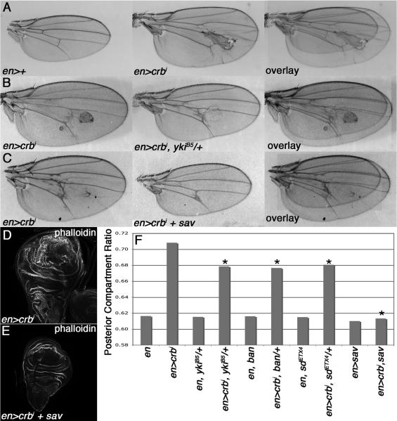
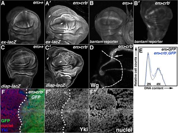
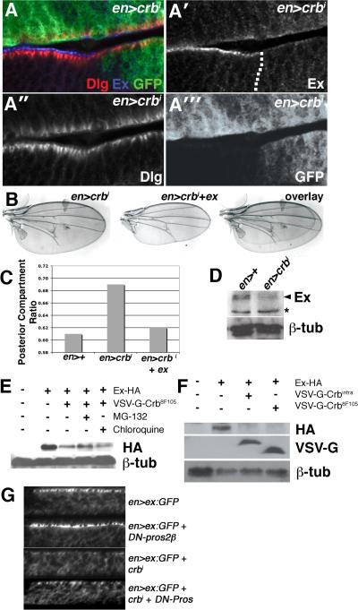
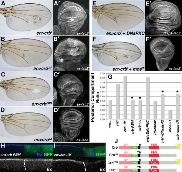
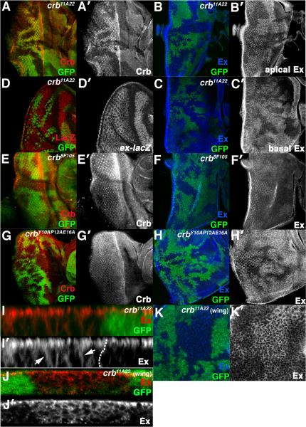
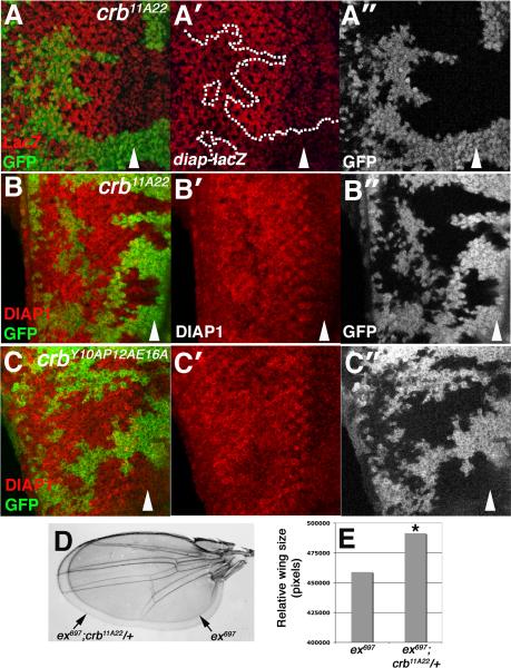
Results: We find that Crb-driven growth is dependent upon the Salvador/Warts/Hippo (SWH) pathway and its transcriptional effector Yorkie (Yki). Expression of the Crb intracellular domain elevates Yki activity, and this correlates in tissues and cultured cells with loss of Expanded (Ex), an apically localized SWH component that inhibits Yki. Reciprocally, loss of crb elevates Ex levels, although this excess Ex does not concentrate to its normal location at apical junctions. The Ex-regulatory domain of Crb maps to the juxtamembrane FERM-binding motif (JM), a cytoskeletal interaction domain distinct from the PDZ-binding motif (PBM) through which Crb binds polarity factors. Expression of Crb-JM drives Yki activity and organ growth with little effect on tissue architecture, while Crb-PBM reciprocally produces tissue architectural defects without significant effect on Yki activity.

Conclusions: These studies identify Crb as a novel SWH regulator via JM-dependent effects on Ex levels and localization and suggest that discrete domains within Crb may allow it to integrate junctional polarity signals with a conserved growth pathway.


Figures

Figure 1
Figure 1. Overgrowth driven by the crbi transgene is sensitive to the dose of SWH pathway genes
Images and overlays of (A) transgenic en>crbi and control en>+, (B) en>crbi and en>crbi,ykiB5/+, (C) en>crbi and en>crbi,sav wings. (D-E) Phalloidin-FITC staining of en>crbi and en>crbi,sav larval wings. (F) PCR in the indicated genotypes (minimum 10 wings per genotype; * p<0.05 compared to en>crbi wings).
Figure 2
Figure 2. crbi elevates Yki-activity
α-β-gal staining or GFP fluorescence in wing discs carrying (A) ex-lacZ, (B) ban-GFP, or (C) diap1-lacZ in the background of (A,B,C) en>+ or (A′,B′,C′) en>crbi. Arrows in A′ and C′ highlight elevated ex-lacZ and diap1-lacZ expression. (D) α-Wg stain in en>crbi wings discs (posterior = right of dashed line). (E) FACS-analysis of en>GFP (black) and transgenic en>crbi,GFP (blue) wing discs. (F) Co-staining for Yki (blue) and HP1 (nuclei; red) in en>crbi,GFP discs (posterior = right of dotted line).
Figure 3
Figure 3. crbi downregulates Ex levels
(A-A′″) Lateral section of en>crbi,GFP wing disc co-stained for Dlg (red) and Ex (blue). Dotted line denotes A:P boundary. (B) Images and overlays of en>crbi and en>crbi,ex wings. (C) PCR in the indicated genotypes. (D) Immunoblot of Ex in en>+ and en>crbi wing discs. Arrowhead denotes Ex based on comigration with overexpressed Ex (not shown) (* = non-specific band). Lower panel is α-β-tub loading control. (E) Immunoblot of HA-Ex in Crb8F105-expressing cells treated with the MG132 (lane 4) or chloroquine (lane 5). (F) Corresponding α-HA, α-VSV-G, and α-β-tub immunoblots of S2 cells expressing HA-Ex from the pAct-HA-Ex plasmid (lanes 2-4), and VSV-G-tagged forms of either crbi (lane 3) or crb8F105 (lane 4) from the pMT plasmid. (G) Lateral images of Ex:GFP in the posterior region of the wing pouch in the indicated genotypes.
Figure 4
Figure 4. The Crb-JM controls Ex levels and Yki activity
Paired light microscopic (A-F) and confocal images of α-β-gal staining to detect activity of the ex-lacZ transgene (A′-D′,F′) or diap1-lacZ (E′) in the indicated genotypes. (G) PCR values in the indicated genotypes. (*p<0.05 compared to en>crbi). α-Ex (blue) staining in (H) en>crb-PBM,GFP and (I) en>crb-JM,GFP wing discs. Dotted line marks the A:P boundary. (posterior = right). Cartoon of crb transgenes; signal peptide (SP), myc tag (Myc), transmembrane domain (TM), juxtamembrane FERM-binding motif (JM), PDZ-binding motif (PBM), and amino acid substitutions are indicated [adapted from 40].
Figure 5
Figure 5. Effect of crb loss on Ex in disc cells
Confocal images of crb11A22 (A-D, I-K), crb8F105 (E-F), or crbY10AP12AE16A (G-H) clones in the eye (A-I) or wing (J-K) stained for Crb (A,E,G), Ex (B,C,F,H-K). (B) and (C) are apical and basal planes of the same disc. Arrowheads in (I) denote excess Ex in crb11A22 cells that fails to localize apically. Disc in (I) is imaged through apical portion of epithelium; disc in (J) is imaged through entire epithelium. (D) α-β-gal staining to detect activity of the ex-lacZ transgene in crb11A22 eye clones.
Figure 6
Figure 6. crb alleles interact with ex and elevate DIAP1 expression posterior to the furrow
Images of (A-B) crb11A22 or (C) crbY10AP12AE16A clones (lacking GFP) in larval eye discs stained with (A) α-β-Gal to detect diap1-lacZ (red) or (B-C) α-DIAP1 (red). Arrowheads denote position of the MF. (posterior = left). Dotted line in (A) highlights a crb clone that projects posterior to the MF and expresses elevated diap1-lacZ. (D) Optical overlay and (E) size adult wings of the indicated genotypes. (* p<1.5×10⁻⁷ relative to ex697 wings).

Comment in

Similar articles

Cited by

References

    1. Bilder D. Epithelial polarity and proliferation control: links from the Drosophila neoplastic tumor suppressors. Genes Dev. 2004;18:1909–1925. - PubMed
    1. Humbert PO, Grzeschik NA, Brumby AM, Galea R, Elsum I, Richardson HE. Control of tumourigenesis by the Scribble/Dlg/Lgl polarity module. Oncogene. 2008;27:6888–6907. - PubMed
    1. Wodarz A, Hinz U, Engelbert M, Knust E. Expression of crumbs confers apical character on plasma membrane domains of ectodermal epithelia of Drosophila. Cell. 1995;82:67–76. - PubMed
    1. Lu H, Bilder D. Endocytic control of epithelial polarity and proliferation in Drosophila. Nat Cell Biol. 2005;7:1132–1139. - PubMed
    1. Moberg KH, Schelble S, Burdick SK, Hariharan IK. Mutations in erupted, the Drosophila ortholog of mammalian tumor susceptibility gene 101, elicit non-cell-autonomous overgrowth. Dev Cell. 2005;9:699–710. - PubMed

Publication types

MeSH terms

Substances

LinkOut - more resources