. 2010 Apr 2;6(4):336-347.
doi: 10.1016/j.stem.2010.03.001.

Efficient derivation of functional floor plate tissue from human embryonic stem cells

Affiliations

Efficient derivation of functional floor plate tissue from human embryonic stem cells

Christopher A Fasano et al. Cell Stem Cell. .

Retraction in

Abstract

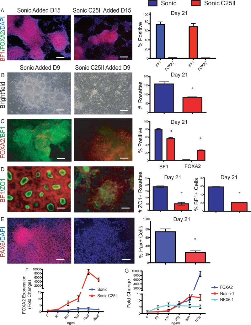
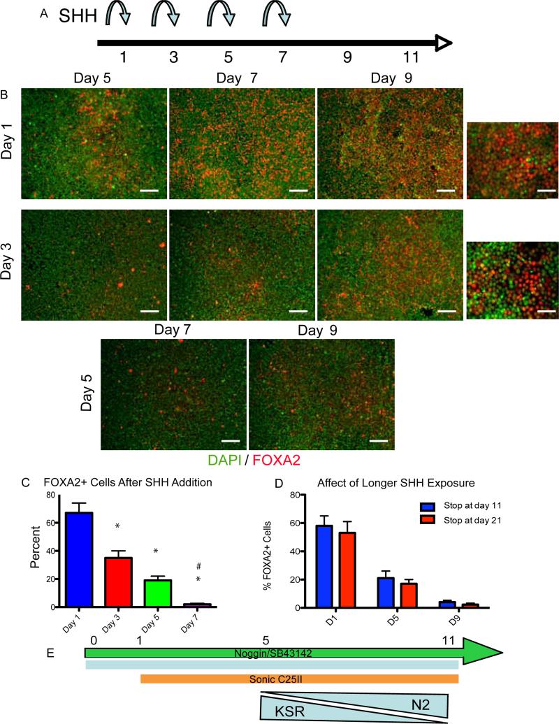
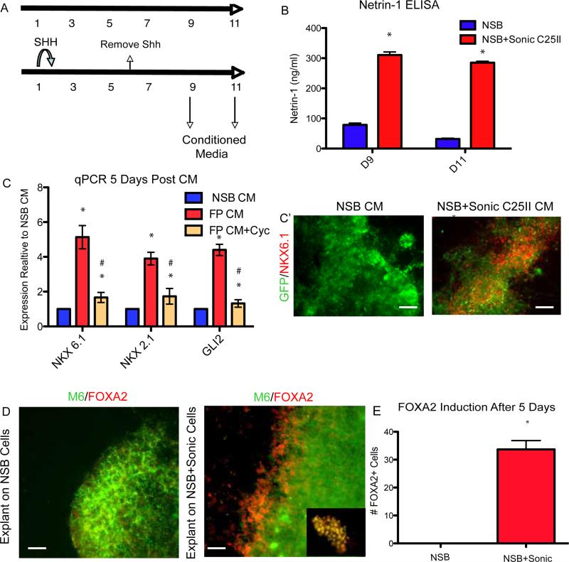
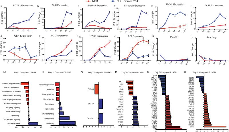
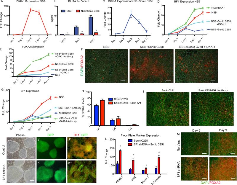
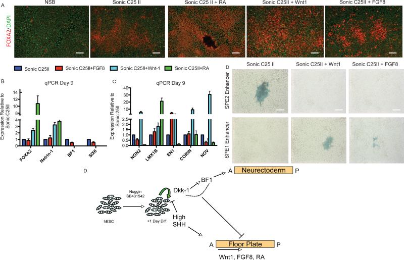
The floor plate (FP) is a critical signaling center during neural development located along the ventral midline of the embryo. Little is known about human FP development because of the lack of tissue accessibility. Here we report the efficient derivation of human embryonic stem cell (hESC)-derived FP tissue capable of secreting Netrin-1 and SHH and patterning primary and hESC derived tissues. FP induction in hESCs is dependent on early SHH exposure and occurs at the expense of anterior neurectoderm (AN). Global gene expression and functional studies identify SHH-mediated inhibition of Dkk-1 as key factor in FP versus AN specification. hESC-derived FP tissue is shown to be of anterior SIX6+ character but is responsive to caudalizing factors suppressing SIX6 expression and inducing a shift in usage of region-specific SHH enhancers. These data define the early signals that drive human FP versus AN specification and determine regional identity in hESC-derived FP.


Figures

Figure 1
Figure 1
High SHH levels increase FOXA2 and decrease BF1 expression (A) Passage 1, Day 21 of neural differentiation shows no effect of SHH treatment when added at Day 15. Results quantified on right, *p<0.01 N=3. Scale bar, 200um. (B) Day 21 of neural differentiation shows a reduction of rosette like structures after Sonic C25II treatment Day 9. Loss of rosettes quantified on right, *p<0.01 N=4. Scale bar, 100um. (C) Sonic C25II treatment results in a decrease of BF1 and an increase in Foxa2 at Day 21. Quantified on right, *p<0.05 N=4. Scale bar, 200um. (D) Day 21 of neural differentiation reveals a decrease in ZO1/BF1+ rosette structures. This decrease is quantified, *p<0.01 N=4. Scale bar, 50um. (E) Decrease in PAX6 expression at Day 21 after Sonic C25II treatment. This decrease is quantified, *p<0.01 N=4. Scale bar, 200um. (F) Dose response curve comparing Sonic and Sonic C25II efficacy on FOXA2 induction. (E) Dose response curve of Sonic C25II comparing the induction of FP markers (FOXA2 and Netrin-1) to another SHH responsive gene NKX6.1. See also Figure S1
Figure 2
Figure 2
Floor Plate induction has an early, short temporal patterning window (A) Schematic showing different time points of Sonic C25II additions during neural induction protocol. (B-C) Heading on the left delineates the day Sonic C25II was added, heading on the top delineates when the assay was stopped. The earlier Sonic C25II is added, and the longer the cells are exposed to it, leads to very high percentages of FOXA2. (C) This result is quantified, *p<0.01 N=3. Scale bars, 200um, high mag, 50um. (D) Extended treatment with Sonic C25II (9 days of exposure) does not yield increased FOXA2 induction. (E) Schematic of optimal protocol for FOXA2 induction to be used for the rest of the study.
Figure 3
Figure 3
hESC derived FP is functional (A) Schematic showing when conditioned media was collected. (B) ELISA showing an increase in levels of Netrin-1 secreted into the media at Days 9 and 11 when Sonic C25II is added early to the neural induction, *p<0.01 N=3. (C) Conditioned media from NSB and NSB+Sonic C25II was collected and placed on cultures containing NSB derived neural precursor cells qRT-PCR showing an induction of ventral genes (NKX6.1 and NKX2.1) as well as the SHH responsive gene (GLI2). These inductions are repressed in the presence of the SHH antagonist cyclopamine. (C’) The induction of NKX6.1 is shown at the level of the protein using a GFP expressing line. *p<0.01 compared to NSB CM, #p<0.05 compared to FP CM, N=3. Scale bar, 200um. (D and E) Neural explants isolated from E8.5 neurectoderm co-cultured with NSB+Sonic C25II tissue show ectopic FOXA2 staining. Inset shows co-localization of M6 (Green) and FOXA2 (Red). (E) This data is quantified, *p<0.001 N=4 explants. Scale bar, 50um. See also Figure S3
Figure 4
Figure 4
Detailed transcriptional analysis reveals novel genes involved in FP development. (A-J) qRT-PCR data showing time course of expression over the length of the 11 day protocol. The genes looked at represented different populations including FP markers (A-D), SHH responsive genes (E-G), neural markers (H), AN markers (I and J), and genes involved in mesodermal and endodermal commitment (K and L). (M-R) Detailed time course microarray analysis (M-N) GO terms for Day 7 (M) and Day 11 (N) showing increase or decrease compared to NSB control. FP condition shows enrichment in genes associated with axon guidance and secreted proteins, while showing a decrease in genes associated with anterior neurectoderm development. (O-R) Pair wise comparisons showing genes up and down regulated compared to NSB control condition at Day 3 (O), Day 5 (P), Day 7 (Q), and Day 11 (R). See also Figure S3 and Tables S1-18
Figure 5
Figure 5
DKK-1 inhibits FP induction (A) qPCR for DKK-1 expression in control NSB condition over time. (B) ELISA measuring DKK-1 protein levels in the media at Day 5, 7, and 11 showing a decrease in Dkk-1 levels after Sonic C25II treatment, *p<0.05 N=3. (C) qPCR for DKK-1 expression in NSB+Sonic C25II condition over time. (D and E) qPCR for BF1 (D) and FOXA2 (E) showing an increase in BF1 and decrease of FOXA2 after DKK-1 addition, and an increase in FOXA2 when DKK-1 antibody is added. (F) Immunostaining for FOXA2 showing a decrease in FOXA2+ cells when DKK-1 is added. Scale bar, 200um. (G) qPCR for BF1 expression showing that DKK-1 antibody treatment leads to a decreased expression at earlier time points (Day 3-Day 5). (H and I) Early addition of DKK-1 antibody leads to an increase of FOXA2 expression, but has no effect when added at later timepoints. (I) Immunocytochemical data demonstrating that Dkk-1 treatment starting at day 5 of differentiation (or later) does not enhance SHH-mediated FOXA2 expression. (J-K”) hESC transduced with either control or BF1 shRNA (J and K), GFP is a marker of transduction (A’ and B’). When differentiated to neural tissue, a reduction of BF1 is seen at the level of the protein compared to control (J” and K”). Scale bars, A and B 100um, J” and K” 200um. (L) qRT-PCR analysis at Day 11 showed an increase in FP markers (FOXA2, SHH, Netrin-1 and F-Spondin) in the BF1 shRNA line compared to the control, p<0.01 N=3. (M) BF1 shRNA leads to an upregulation of FOXA2 seen at the level of the protein. Scale bar, 200um. See also Figure S4
Figure 6
Figure 6
hESC derived FP can be shifted along the A/P axis (A) Immunostaining reveals an increase in FOXA2 in response to FGF8, Wnt-1, and Retinoic Acid. Scale bar, 200um. (B) qPCR showing caudilizing agents such as FGF8, Wnt-1, and Retinoic Acid (RA) lead to an increase in FOXA2 and a reduction in SIX6 compared to NSB+Sonic C25II. (C) qPCR for a panel of midbrain FP markers (CORIN and NOV) and midbrain DA progenitor markers (LMX1B, NGN2, and EN1). (D) FP cells were transfected with a SHH enhancer that drives expression to the anterior ventral axis (SBE2) or midbrain ventral axis (SBE1). The default FP exhibits SBE2 activity indicating an anterior location. This is abolished upon Wnt1 and FGF8 addition and SBE1 activity is now seen suggesting a shift from anterior identity to midbrain. Scale bar, 200um. (E) Schematic of FP versus AN specification during hESC differentiation. Neural differentiation is initiated upon exposure to Noggin and SB431542. SHH exposure, starting at day 1 of differentiation, induces FP differentiation and via inhibition of DKK-1 and BF1 suppresses AN specification. The regional identity of the resulting FP cells is anterior by default but posterior FP tissue can be induced in the presence of caudalizing factors such as Wnt-1, FGFF8 or RA. See also Figure S5

References

    1. Barberi T, Klivenyi P, Calingasan NY, Lee H, Kawamata H, Loonam K, Perrier AL, Bruses J, Rubio ME, Topf N, Tabar V, Harrison NL, Beal MF, Moore MA, Studer L. Neural subtype specification of fertilization and nuclear transfer embryonic stem cells and application in parkinsonian mice. Nat Biotechnol. 2003;10:1200–1207. - PubMed
    1. Briscoe J, Ericson J. The specification of neuronal identity by graded Sonic Hedgehog signaling. Semin Cell Dev Biol. 1999;3:353–62. - PubMed
    1. Burns CJ, Zhang J, Brown EC, Van Bibber AM, Van Es J, Clevers H, Ishikawa TO, Taketo MM, Vetter ML, Fuhrmann S. Investigation of Frizzled-5 during neural development in mouse. Dev Dyn. 2008;237:1614–1626. - PMC - PubMed
    1. Chambers SM, Fasano CA, Papapetrou EP, Tomishima M, Sadelain M, Studer L. Highly efficient neural conversion of human ES and iPS cells by dual inhibition of SMAD signaling. Nat Biotechnol. 2009;27:275–280. - PMC - PubMed
    1. Charrier JB, Lapointe F, Le Douarin NM, Teillet MA. Dual origin of the floor plate in the avian embryo. Development. 2002;129:4785–4796. - PubMed

Publication types

MeSH terms