Recognition of decay accelerating factor and alpha(v)beta(3) by inactivated hantaviruses: Toward the development of high-throughput screening flow cytometry assays
- PMID: 20363206
- PMCID: PMC2905740
- DOI: 10.1016/j.ab.2010.03.016
Recognition of decay accelerating factor and alpha(v)beta(3) by inactivated hantaviruses: Toward the development of high-throughput screening flow cytometry assays
Abstract
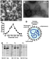
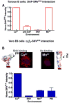
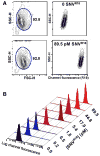
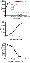
Hantaviruses cause two severe diseases in humans: hemorrhagic fever with renal syndrome (HFRS) and hantavirus cardiopulmonary syndrome (HCPS). The lack of vaccines or specific drugs to prevent or treat HFRS and HCPS and the requirement for conducting experiments in a biosafety level 3 laboratory (BSL-3) limit the ability to probe the mechanism of infection and disease pathogenesis. In this study, we developed a generalizable spectroscopic assay to quantify saturable fluorophore sites solubilized in envelope membranes of Sin Nombre virus (SNV) particles. We then used flow cytometry and live cell confocal fluorescence microscopy imaging to show that ultraviolet (UV)-killed SNV particles bind to the cognate receptors of live virions, namely, decay accelerating factor (DAF/CD55) expressed on Tanoue B cells and alpha(v)beta(3) integrins expressed on Vero E6 cells. SNV binding to DAF is multivalent and of high affinity (K(d) approximately 26pM). Self-exchange competition binding assays between fluorescently labeled SNV and unlabeled SNV are used to evaluate an infectious unit-to-particle ratio of approximately 1:14,000. We configured the assay for measuring the binding of fluorescently labeled SNV to Tanoue B suspension cells using a high-throughput flow cytometer. In this way, we established a proof-of-principle high-throughput screening (HTS) assay for binding inhibition. This is a first step toward developing HTS format assays for small molecule inhibitors of viral-cell interactions as well as dissecting the mechanism of infection in a BSL-2 environment.
2010 Elsevier Inc. All rights reserved.
Figures


Similar articles
-
A High-Throughput Flow Cytometry Screen Identifies Molecules That Inhibit Hantavirus Cell Entry.SLAS Discov. 2018 Aug;23(7):634-645. doi: 10.1177/2472555218766623. Epub 2018 Apr 2. SLAS Discov. 2018. PMID: 29608398 Free PMC article.
-
Equilibrium and kinetics of Sin Nombre hantavirus binding at DAF/CD55 functionalized bead surfaces.Viruses. 2014 Mar 10;6(3):1091-111. doi: 10.3390/v6031091. Viruses. 2014. PMID: 24618810 Free PMC article.
-
A novel Sin Nombre virus DNA vaccine and its inclusion in a candidate pan-hantavirus vaccine against hantavirus pulmonary syndrome (HPS) and hemorrhagic fever with renal syndrome (HFRS).Vaccine. 2013 Sep 13;31(40):4314-21. doi: 10.1016/j.vaccine.2013.07.025. Epub 2013 Jul 24. Vaccine. 2013. PMID: 23892100 Free PMC article.
-
Hantavirus Cardiopulmonary Syndrome in Canada.Emerg Infect Dis. 2020 Dec;26(12):3020-3024. doi: 10.3201/eid2612.202808. Emerg Infect Dis. 2020. PMID: 33219792 Free PMC article. Review.
-
Hantavirus pulmonary syndrome.Virus Res. 2011 Dec;162(1-2):138-47. doi: 10.1016/j.virusres.2011.09.017. Epub 2011 Sep 17. Virus Res. 2011. PMID: 21945215 Review.
Cited by
-
Protocadherin-1 is essential for cell entry by New World hantaviruses.Nature. 2018 Nov;563(7732):559-563. doi: 10.1038/s41586-018-0702-1. Epub 2018 Nov 21. Nature. 2018. PMID: 30464266 Free PMC article.
-
Vascular dysfunction in hemorrhagic viral fevers: opportunities for organotypic modeling.Biofabrication. 2024 Jun 5;16(3):032008. doi: 10.1088/1758-5090/ad4c0b. Biofabrication. 2024. PMID: 38749416 Free PMC article. Review.
-
Rapid parallel flow cytometry assays of active GTPases using effector beads.Anal Biochem. 2013 Nov 15;442(2):149-57. doi: 10.1016/j.ab.2013.07.039. Epub 2013 Aug 6. Anal Biochem. 2013. PMID: 23928044 Free PMC article.
-
Pathogenic old world hantaviruses infect renal glomerular and tubular cells and induce disassembling of cell-to-cell contacts.J Virol. 2011 Oct;85(19):9811-23. doi: 10.1128/JVI.00568-11. Epub 2011 Jul 20. J Virol. 2011. PMID: 21775443 Free PMC article.
-
Replication kinetics of pathogenic Eurasian orthohantaviruses in human mesangial cells.Virol J. 2024 Oct 1;21(1):241. doi: 10.1186/s12985-024-02517-5. Virol J. 2024. PMID: 39354507 Free PMC article.
References
-
- Hjelle B, Anderson B, Torrez-Martinez N, Song W, Gannon WL, Yates TL. Prevalence and geographic genetic variation of hantaviruses of New World harvest mice (Reithrodontomys): identification of a divergent genotype from a Costa Rican Reithrodontomys mexicanus. Virology. 1995;207:452–459. - PubMed
-
- Hjelle B, Jenison SA, Goade DE, Green WB, Feddersen RM, Scott AA. Hantaviruses: clinical, microbiologic, and epidemiologic aspects. Crit Rev Clin Lab Sci. 1995;32:469–508. - PubMed
-
- Jonsson CB, Schmaljohn CS. Replication of hantaviruses. Curr Top Microbiol Immunol. 2001;256:15–32. - PubMed
-
- Jonsson CB, Milligan BG, Arterburn JB. Potential importance of error catastrophe to the development of antiviral strategies for hantaviruses. Virus Res. 2005;107:195–205. - PubMed
Publication types
MeSH terms
Substances
Grants and funding
- K25AI060036/AI/NIAID NIH HHS/United States
- U54 MH084690/MH/NIMH NIH HHS/United States
- UO1AI56618/AI/NIAID NIH HHS/United States
- U01 AI056618/AI/NIAID NIH HHS/United States
- U54MH084690/MH/NIMH NIH HHS/United States
- P30CA118100/CA/NCI NIH HHS/United States
- K25 AI060036/AI/NIAID NIH HHS/United States
- U54MH074425/MH/NIMH NIH HHS/United States
- UO1AI054779/AI/NIAID NIH HHS/United States
- U54 MH074425/MH/NIMH NIH HHS/United States
- P30 CA118100/CA/NCI NIH HHS/United States
- F32 AI074246/AI/NIAID NIH HHS/United States
- U01 AI054779/AI/NIAID NIH HHS/United States
LinkOut - more resources
Full Text Sources
Other Literature Sources
Miscellaneous

