. 2010 May 21;475(3):174-8.
doi: 10.1016/j.neulet.2010.03.073. Epub 2010 Apr 2.

HIV-1 protein-mediated amyloidogenesis in rat hippocampal cell cultures

Affiliations

HIV-1 protein-mediated amyloidogenesis in rat hippocampal cell cultures

M Y Aksenov et al. Neurosci Lett. .

Abstract

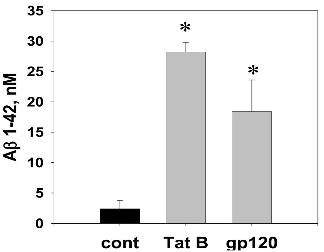
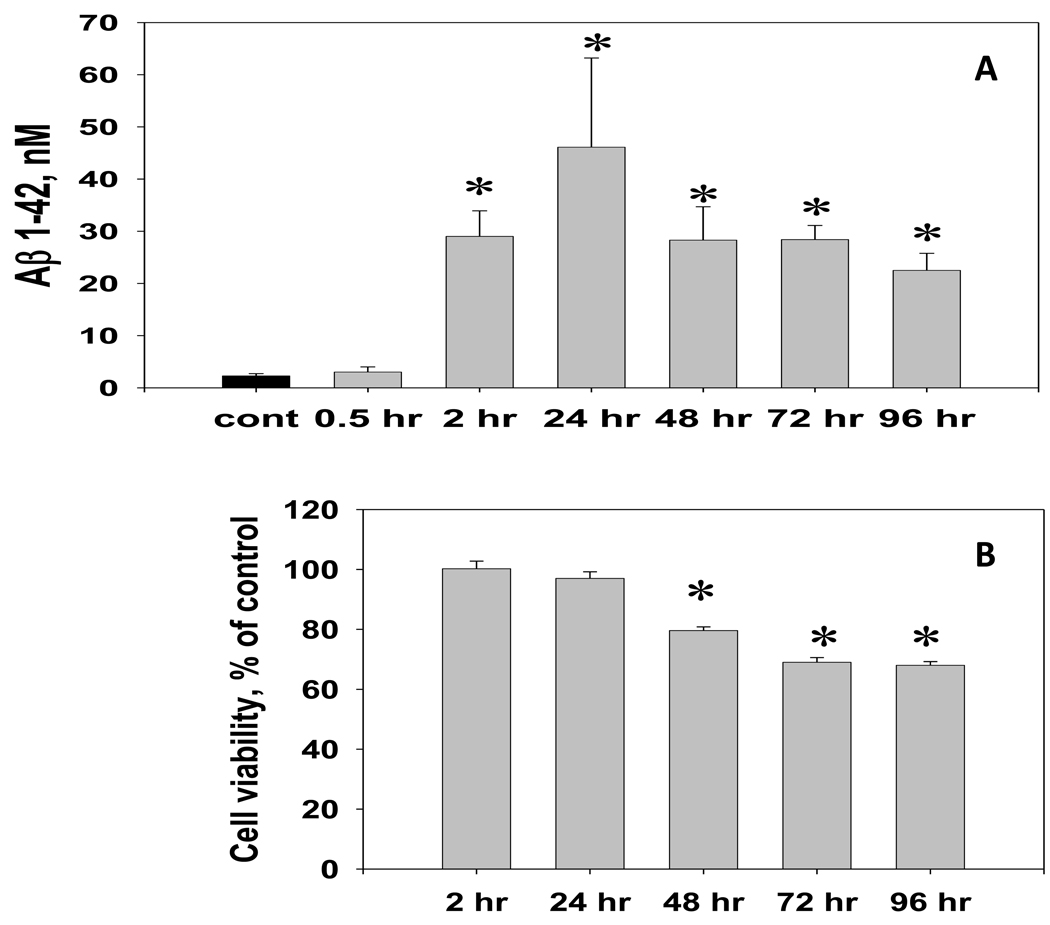
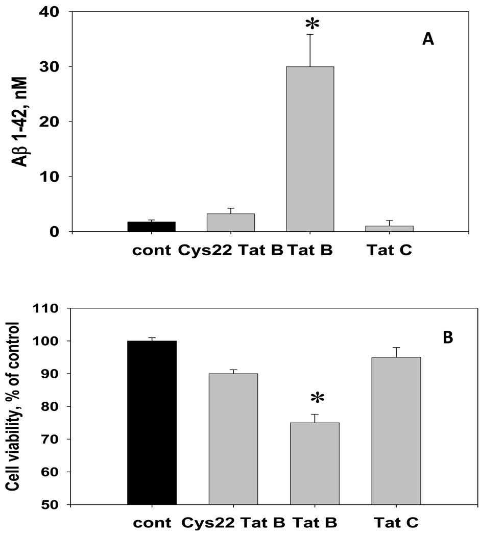
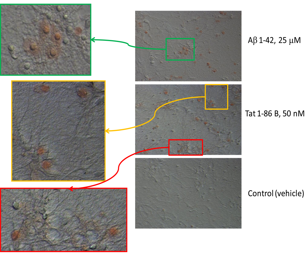
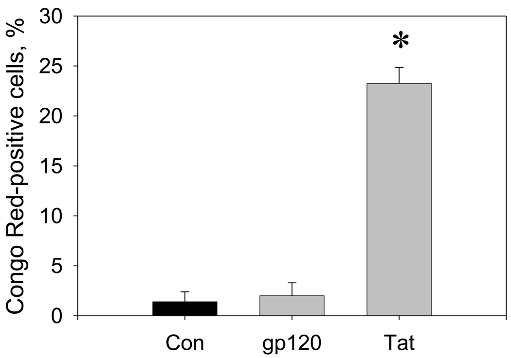
Since the beginning of the highly active antiretroviral therapy (HAART) era, epidemiological evidence indicates an increasing incidence of Alzheimer's (AD)-like brain pathology in aging HIV patients. Emerging evidence warns of potential convergent mechanisms underlying HIV- and Abeta-mediated neurodegeneration. We found that HIV-1 Tat B and gp120 promote the secretion of Abeta 1-42 in primary rat fetal hippocampal cell cultures. Our results demonstrate that the variant of Tat expressed by the neurotropic subtype of HIV-1 virus (HIV-1 clade B) specifically induces both the release of amyloidogenic Abeta 1-42 and the accumulation of cell-bound amyloid aggregates. The results of the research rationalize testing of the ability of beta-amyloid aggregation inhibitors to attenuate HIV protein-mediated cognitive deficits in animal models of NeuroAIDS. The long-term goal of the study is to evaluate the potential benefits of anti-amyloidogenic therapies for management of cognitive dysfunction in aging HIV-1 patients.


Figures

Figure 1
Figure 1. The Aβ 1–42 immunoreactivity in the conditioned medium from hippocampal cell cultures exposed to HIV-1 Tat or gp120
The graph represents results of direct anti-Aβ 1–42 ELISA measurements in the CM samples collected from hippocampal cultures (n=8 per group) treated with either 100 nM Tat 1–86 B, 100 nM gp120, or the equal volume of vehicle. The freshly prepared Aβ 1–42 stock was serially diluted with cell culture growth medium to obtain ELISA calibration curves. Data presented as mean values ± SEM. *- marks significant (P<0.05) differences in extracellular Aβ 1–42 immunoreactivity.
Figure 2
Figure 2. The temporal relationship between the Aβ 1–42 release and cell viability changes in hippocampal cell cultures exposed to a toxic dose of Tat 1–86 B
A. The Aβ 1–42 immunoreactivity was measured in CM samples collected from hippocampal cultures during the continuous exposure (0.5 – 96 hours) to 50 nM Tat 1–86 or vehicle (n=5 per group). Data are presented as mean values ± SEM. *- marks significant (P<0.05) differences in extracellular Aβ 1–42 immunoreactivity between Tat-treated and vehicle-treated cell cultures. B. Cell viability in hippocampal cell cultures (treated side-by-side, n=8 per group) was determined using Live/Dead assay. Data are presented as mean % of control values ± SEM. *- marks incubation time points for significant (P<0.05) differences in Live/Dead ratios between cultures treated with 50 nM Tat 1–86 and vehicle-treated controls have been observed.
Figure 3
Figure 3. The effect of non-neurotoxic variants of HIV-1 Tat (Cys22 Tat 1–86 B and Tat 1–101 C) on the Aβ 1–42 production in hippocampal cell cultures
The graph represents results of direct anti-Aβ 1–42 ELISA measurements in the CM samples collected from individual cultures (n=8 per group) treated with 100 nM Tat 1–86 B, Cys22 Tat 1–86 B, Tat 1–101 C, or the equal volume of vehicle for 72 hours. Data are presented as mean values ± SEM. Cell viability in hippocampal cell cultures was determined using Live/Dead assay. *- marks significant (P<0.05) differences in hippocampal cell viability or extracellular Aβ 1–42 immunoreactivity.
Figure 4
Figure 4. The detection of cell-bound β-amyloid aggregates by the Congo Red staining in living hippocampal cells exposed to Aβ 1–42 or HIV-1 Tat 1–86 B
Representative images show the Congo Red binding to hippocampal cell cultures exposed to a toxic dose of Aβ 1–42 (40-hour exposure), Tat 1–86 B (24-hour exposure), or vehicle. Results of the Congo Red staining in cultures treated with 100 nM gp120 for 72 hours, 100 nM Cys22 Tat or Tat 1–101 C for 24 hours were not different from the vehicle-treated controls and are not shown in the Figure. Colored boxes mark areas of Congo Red/DIC images, which are shown as new magnified images with greater resolution.
Figure 5
Figure 5. Changes in numbers of Congo Red-positive cells in hippocampal cell cultures exposed to HIV-1 Tat or gp120
The graph represents relative numbers (% of total cells) of Congo Red-positive cells in hippocampal cell cultures after 3 days of treatment with vehicle (control), gp120 (100 nM), or Tat 1–86 B (100 nM). For each individual cell culture in a group (n=4), Congo Red-positive hippocampal cells were counted in 4 random fields of vision. For each field of vision (0.5×0.37 mm) results were normalized to the subsequent total cell number determined using Hoechst staining of cell nuclei (typically 70–95 cells per field). Data are presented as mean values ± SEM. *- marks significant (P<0.05) differences in relative numbers of Congo Red-positive cells.

Similar articles

Cited by

References

    1. Aksenov MY, Aksenova MV, Carney JM, Butterfield DA. Alpha 1-antichymotrypsin interaction with A beta (1–42) does not inhibit fibril formation but attenuates the peptide toxicity. Neurosci Lett. 1996;217:117–120. - PubMed
    1. Aksenov MY, Aksenova MV, Nath A, Ray PD, Mactutus CF, Booze RM. Cocaine-mediated enhancement of Tat toxicity in rat hippocampal cell cultures: the role of oxidative stress and D1 dopamine receptor. Neurotoxicology. 2006;27:217–228. - PubMed
    1. Aksenov MY, Aksenova MV, Mactutus CF, Booze RM. Attenuated neurotoxicity of the transactivation-defective HIV-1 Tat protein in hippocampal cell cultures. Exp Neurol. 2009;219:586–590. - PMC - PubMed
    1. Aksenova MV, Aksenov MY, Adams SM, Mactutus CF, Booze RM. Neuronal survival and resistance to HIV-1 Tat toxicity in the primary culture of rat fetal neurons. Exp Neurol. 2009;215:253–263. - PMC - PubMed
    1. Apcher GS, Heink S, Zantopf D, Kloetzel PM, Schmid HP, Mayer RJ, Krüger E. Human immunodeficiency virus-1 Tat protein interacts with distinct proteasomal alpha and beta subunits. FEBS Lett. 2003;553:200–204. - PubMed

Publication types

MeSH terms

Substances

LinkOut - more resources