. 2010 Jun 16;168(1):263-72.
doi: 10.1016/j.neuroscience.2010.03.049. Epub 2010 Apr 2.

The parabrachial nucleus is a critical link in the transmission of short latency nociceptive information to midbrain dopaminergic neurons

Affiliations

The parabrachial nucleus is a critical link in the transmission of short latency nociceptive information to midbrain dopaminergic neurons

V Coizet et al. Neuroscience. .

Abstract

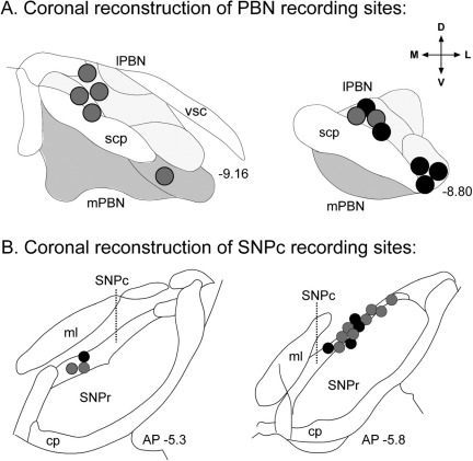
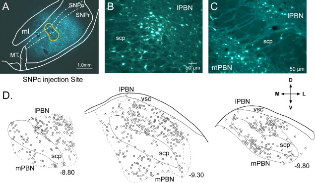
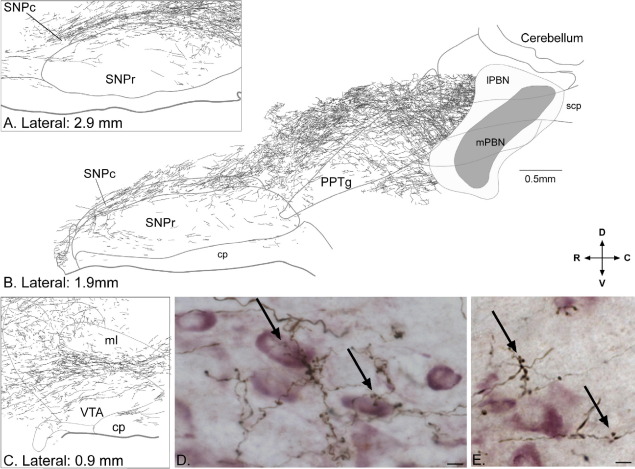
Many dopaminergic neurons exhibit a short-latency response to noxious stimuli, the source of which is unknown. Here we report that the nociceptive-recipient parabrachial nucleus appears to be a critical link in the transmission of pain related information to dopaminergic neurons. Injections of retrograde tracer into the substantia nigra pars compacta of the rat labelled neurons in both the lateral and medial parts of the parabrachial nucleus, and intra-parabrachial injections of anterograde tracers revealed robust projections to the pars compacta and ventral tegmental area. Axonal boutons were seen in close association with tyrosine hydroxylase-positive (presumed dopaminergic) and negative elements in these regions. Simultaneous extracellular recordings were made from parabrachial and dopaminergic neurons in the anaesthetized rat, during the application of noxious footshock. Parabrachial neurons exhibited a short-latency, short duration excitation to footshock while dopaminergic neurons exhibited a short-latency inhibition. Response latencies of dopaminergic neurons were reliably longer than those of parabrachial neurons. Intra-parabrachial injections of the local anaesthetic lidocaine or the GABA(A) receptor antagonist muscimol reduced tonic parabrachial activity and the amplitude (and in the case of lidocaine, duration) of the phasic response to footshock. Suppression of parabrachial activity with lidocaine reduced the baseline firing rate of dopaminergic neurons, while both lidocaine and muscimol reduced the amplitude of the phasic inhibitory response to footshock, in the case of lidocaine sometimes abolishing it altogether. Considered together, these results suggest that the parabrachial nucleus is an important source of short-latency nociceptive input to the dopaminergic neurons.


Figures

Suppl. Fig. 1
Suppl. Fig. 1
Fig. 1
Fig. 1
Parabrachial neurons retrogradely labelled from the substantia nigra pars compacta (SNPc) are found in both the lateral and medial parts of the parabrachial nucleus (PBN). (A) Photomicrograph of a fluorogold (retrograde tracer) injection site in the SNPc. (B) Photo-micrograph of retrogradely labelled neurons in the lateral parabrachial nucleus (lPBN) and (C) lateral and medial parabrachial nucleus (mPBN). (D) Quantitative plot of labelled neurons at different anterior-posterior levels of the PBN (numbers associated with each section indicate its location caudal to bregma in mm, according to the atlas of Paxinos and Watson, 1998). The directional arrows apply to (A–D); M, medial; L, lateral; D, dorsal; V, ventral. Additional abbreviations: ml, medial lemniscus; MT, medial terminal nucleus of the accessory optic tract; scp, superior cerebellar peduncle; SNPr, substantia nigra pars reticulata; vsc, ventral spino–cerebellar tract.
Fig. 2
Fig. 2
A robust projection from the parabrachial nucleus (PBN) to the substantia nigra pars compacta (SNPc) and ventral tegmental area (VTA) was evident. (A–C) Drawings based on photo-micrographs of parasagittal sections of rat brain following a large injection of the anterograde tracer Phaseolus vulgaris leucoagglutinin (PHA-L) in the PBN (shaded areas in the PBN in B). Dense labelling of fibres and terminal boutons can be seen in the SNPc (A, B) as well as in the VTA (C). (D, E). Examples of terminals and boutons anterogradely labelled from the PBN in relation to TH+ (presumed dopaminergic) neurons in the SNPc. Arrows indicate clusters of terminal boutons in close association with TH+ elements (D) and aggregating away from TH+ elements (E). Scale bar in (B) applies to (A–C) while scale bars in photomicrographs (D, E)=10 μm. In (A–C), laterality of the section is shown in mm according to the atlas of Paxinos and Watson (1998). The directional arrows apply to (A–E); R, rostral; C, caudal; D, dorsal; V, ventral. Additional abbreviations: cp, cerebral peduncle; lPBN, lateral parabrachial nucleus; ml, medial lemniscus; mPBN, medial parabrachial nucleus; PPTg, pedunculopontine tegmental nucleus; SNPr, substantia nigra pars reticulata; scp, superior cerebellar peduncle.
Fig. 3
Fig. 3
Effects of local intraparabrachial injections of lidiocaine on footshock-evoked multi-unit responses in the parabrachial nucleus (PBN) and in a single dopaminergic (DA) neuron. The graphs present raster displays and peri-stimulus histograms of single case data aligned on the presentation of 120 stimuli (0.5 Hz; vertical dotted line; stimulus artifacts have been removed for clarity—these did not overlap with responses in the PBN or in DA neurons). Prior to the injection of lidocaine, both the PBN and the DA neuron (A, B) were responsive to the footshock. Following the injection of lidocaine into the PBN, local neurons became unresponsive to the footshock (C) and so did the DA neuron (D).

References

    1. Benabid A.L., Jeaugey L. Cells of the rat lateral habenula respond to high-threshold somatosensory inputs. Neurosci Lett. 1989;96:289–294. - PubMed
    1. Besson J.M., Chaouch A. Peripheral and spinal mechanisms of nociception. Physiol Rev. 1987;67:67–186. - PubMed
    1. Boksem M.A., Tops M., Kostermans E., De Cremer D. Sensitivity to punishment and reward omission: evidence from error-related ERP components. Biol Psychol. 2008;79:185–192. - PubMed
    1. Brischoux F., Chakraborty S., Brierley D.I., Ungless M.A. Phasic excitation of dopamine neurons in ventral VTA by noxious stimuli. Proc Natl Acad Sci U S A. 2009;106:4894–4899. - PMC - PubMed
    1. Bullitt E. Expression of c-fos-like protein as a marker for neuronal activity following noxious stimulation in the rat. J Comp Neurol. 1990;296:517–530. - PubMed

Publication types