. 2010 Apr;22(4):1174-89.
doi: 10.1105/tpc.109.066647. Epub 2010 Apr 2.

Identification of specific DNA binding residues in the TCP family of transcription factors in Arabidopsis

Affiliations

Identification of specific DNA binding residues in the TCP family of transcription factors in Arabidopsis

Pooja Aggarwal et al. Plant Cell. 2010 Apr.

Abstract

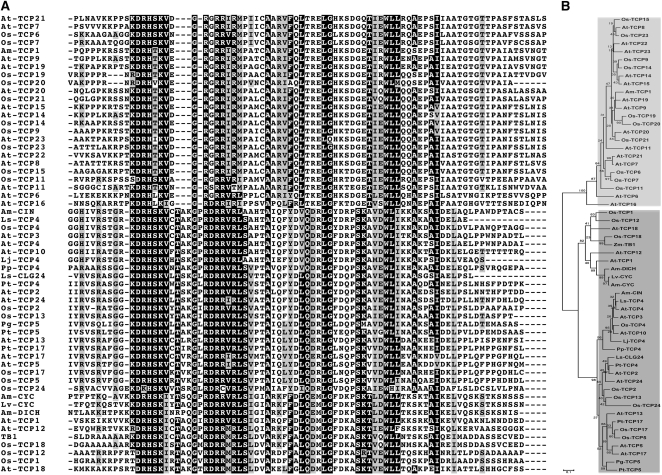
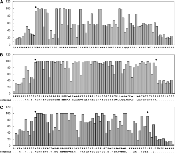
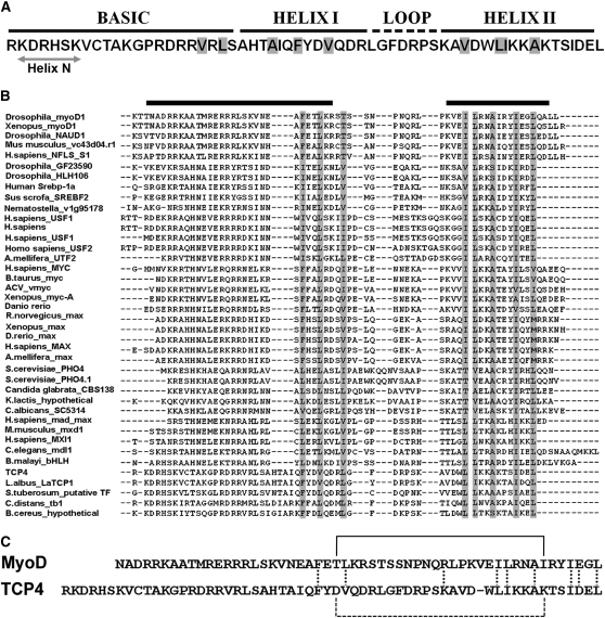
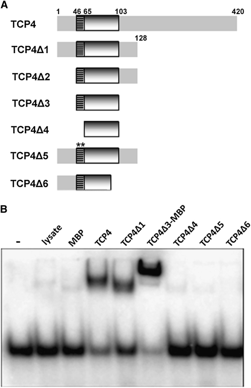
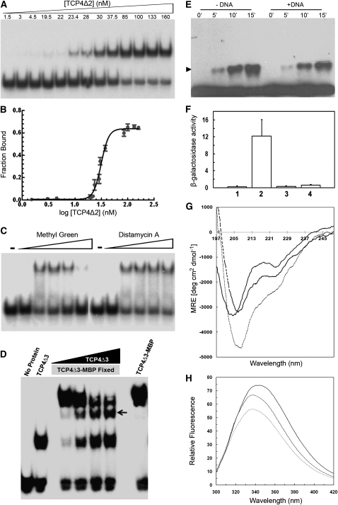
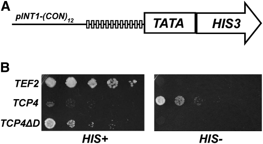
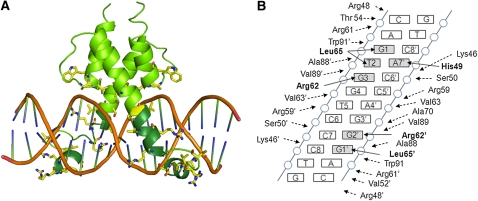
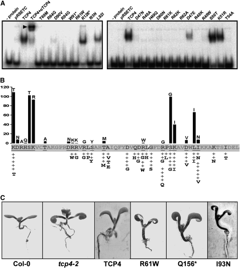
The TCP transcription factors control multiple developmental traits in diverse plant species. Members of this family share an approximately 60-residue-long TCP domain that binds to DNA. The TCP domain is predicted to form a basic helix-loop-helix (bHLH) structure but shares little sequence similarity with canonical bHLH domain. This classifies the TCP domain as a novel class of DNA binding domain specific to the plant kingdom. Little is known about how the TCP domain interacts with its target DNA. We report biochemical characterization and DNA binding properties of a TCP member in Arabidopsis thaliana, TCP4. We have shown that the 58-residue domain of TCP4 is essential and sufficient for binding to DNA and possesses DNA binding parameters comparable to canonical bHLH proteins. Using a yeast-based random mutagenesis screen and site-directed mutants, we identified the residues important for DNA binding and dimer formation. Mutants defective in binding and dimerization failed to rescue the phenotype of an Arabidopsis line lacking the endogenous TCP4 activity. By combining structure prediction, functional characterization of the mutants, and molecular modeling, we suggest a possible DNA binding mechanism for this class of transcription factors.


Figures

Figure 1.
Figure 1.
Sequence Analysis of the TCP Domain. (A) Sequence alignment of 80 residues, including the TCP domain from 57 TCP proteins representing all the clades. The class I domain has an extended sequence conservation at the C terminus (not shaded in this alignment). The class II domain has a three-residue insertion in the basic domain that is absent from class I. (B) A neighbor-joining phylogram with midpoint rooting places these two domains in two major and distinct clades: class I (light gray) and class II (dark gray).
Figure 2.
Figure 2.
Amino Acid Conservation in the TCP Domain. The URF values (see Results) are plotted along the y axis, and the amino acid positions of the domain are shown along the x axis. Analysis is performed with 206 TCP domains belonging to both the classes (A), 121 class I domains (B), or 85 class II TCP domains (C). Predicted left and right borders of the domains are marked by circles and diamonds, respectively. The consensus sequences using ≥50% URF value are given below the x axis.
Figure 3.
Figure 3.
Sequence Comparison between TCP and bHLH Domains. (A) Sequence of the TCP4 domain and the predicted bHLH secondary structure are shown. A predicted short helix at the N terminus (helix N) is indicated. The residues predicted to be buried are shaded in gray. (B) TCP4 sequence profile (see Results) was aligned with the profiles generated for the bHLH proteins of known three-dimensional structure. The residues that are fully conserved or substituted for similar amino acids are highlighted in gray. The helical regions for the MyoD structure are shown by black bars on the top of the alignment. (C) Sequence alignment of MyoD and TCP4 DNA binding domains, highlighting the similarities in the C-terminal HLH region. The residue conservation in the HLH domains is shown by vertical dotted lines. van der Waals contact between Thr-33 and Ile-56 of MyoD (solid line) and a probable salt bridge interaction between corresponding residues Asp-75 and Lys-97 in TCP4 (broken line) are shown. No significant sequence similarity is observed in the N-terminal basic region.
Figure 4.
Figure 4.
Delineation of TCP4 DNA Binding Domain. (A) Deletion versions of TCP4 are shown schematically. Numbers denote the amino acid positions on the full-length TCP4 protein. The basic region is shown as a hatched box, and the HLH region is depicted as a shaded box. The G57L;P58L double mutant is indicated by two asterisks. (B) EMSA gel showing the retardation (top bands) of the free oligonucleotide (bottom bands) by TCP4 mutants (bacterial extract was used as the protein source). Oligo alone and bacterial lysate transformed with pRSET-C only are shown by (−) and (lysate), respectively. MBP, maltose binding protein.
Figure 5.
Figure 5.
Biochemical Characterization of the TCP Domain. (A) EMSA gel showing that a fixed amount of radioactively labeled target oligo incubated with increasing amount of purified TCPΔ2 retards progressively increasing amount of oligos. Numbers above the gel indicate concentration of dimer TCPΔ2. (B) Fraction of bound oligo was quantified from (A) and plotted as a function of dimer protein concentration. Analysis of this data by Hill equation yielded an equilibrium dissociation constant (Kd) of 31.3 ± 2.2 nM. (C) EMSA gel performed with purified TCP4Δ1 and target DNA probe in the presence of increasing amount of methyl green or distamycin A. Free oligo in the absence or the presence of 500 μM dye are shown as (−) and (+), respectively. (D) Detection of homodimerization of the TCP4 DNA binding domain by EMSA. A band of intermediate mobility (indicated by an arrow) appeared when purified TCP4Δ3 (7 kD) and TCP4Δ3-MBP (50.2 kD) were incubated together with the 32P-labeled consensus oligos. The intermediate band increased in intensity when increasing amounts of TCP4Δ3 were incubated with a fixed amount of TCP4Δ3-MBP. (E) Immunoblot of purified hexa-His-tagged TCP4Δ1 incubated with 0.01% of glutaraldehyde for increasing time points and probed with anti-(His)6 antibody. The monomeric TCP4 protein is seen here as a continuous band at the base of the denaturing polyacrylamide gel and the dimer band (arrowhead) of increasing intensity formed with increasing time of incubation. Addition of target oligo to the reaction mixture (+DNA) slightly inhibited dimer formation. (F) The strength of protein–protein interaction, as performed by yeast two-hybrid assay, has been shown as β-galactosidase activity. Each experiment was repeated thrice, and the standard errors are shown as error bars along the y axis. The bait in all the assays is TCP4Δ1. Proteins used as prey are no protein (1), TCP4 (2), I93N (3), and I100T (4) mutants. (G) CD spectra of 5 μM purified TCP4Δ2 with 0 μM (solid line), 1 μM (dashed line) and 5 μM (dotted line) target DNA. The spectra are corrected for the contribution of buffer (for solid line) or buffer plus oligo (for dashed and broken lines). (H) Relative fluorescent intensity spectra of 5 μM purified, truncated TCP4 in the absence (solid line) or presence of 1 μM (dashed line) and 5 μM (dotted line) target oligo.
Figure 6.
Figure 6.
Expression of Full-Length TCP4 Protein Inhibits Yeast Growth. (A) Schematic representation of the construct pINT1-(CON)12 with 12 copies of TCP4 consensus sequence (GTGGTCCC, vertical boxes) upstream of a minimal promoter (TATA) and a HIS3 reporter gene. (B) Growth analysis of the yeast strain harboring either no transgene (TEF2), TCP4 gene under TEF2 promoter (TCP4), or TCP4 lacking the binding domain under TEF2 promoter (TCP4ΔD) performed in presence (HIS+) or absence (HIS) of His supplement.
Figure 7.
Figure 7.
Modeling the Three-Dimensional Structure of the TCP4-DNA Complex. (A) Model of TCP4 domain in dimer form bound to the B-form target DNA sequence GTGGTCCC (in orange). The basic domain is shown in dark green, and residues that interact with the DNA backbone are shown in yellow. (B) Summary of the amino acid–DNA interaction in the TCP4 domain. Residues that make direct contacts with DNA bases (gray rectangle) are shown in bold. A, adenine; T, thymidine; G, guanine.
Figure 8.
Figure 8.
DNA Binding of TCP4 Mutants. (A) EMSA gels showing the interaction of various mutants of TCP4 and the target oligo. The names of the mutants are given at the top of the lanes and radiolabeled oligo only is shown as (− protein). Bacterial cell extracts were used as protein source, and the lysate from bacterial culture that contained empty vector is shown as pRSETC. The supershift (arrowhead) of the wild-type TCP4-DNA complex was brought about by adding anti-(His)6 antibody to the reaction mixture. The asterisk indicates truncated protein due to premature stop codon in the gene. (B) The band intensities in (A) were quantified and shown as a bar diagram in (B). The wild-type residue is shown at the base of the bar, whereas the residue it is mutated to is shown at the top. The intensity of the wild-type TCP4-DNA complex band was considered as 100%, and other values are expressed relative to this value. EMSA was performed for only those mutants where the underlined residues in (B) were mutated. The gray-colored residues in (B) indicate that no mutants in those positions were isolated. The activities of the random mutants as assessed in vivo (see Results and Supplemental Table 3 online) are expressed as + signs and the substituted residues are written below it. (C) Arabidopsis seedlings from the wild type (Columbia-0 [Col-0]), TCP4 null mutant (tcp4-2), and various transgenic lines are shown for their epinasty phenotype of cotyledons. Col-0 has flat cotyledons that bend downwards when TCP4 is mutated (tcp4-2). This phenotype can be rescued by introducing a wild-type TCP4. However, a truncated TCP4 with the first 156 amino acids intact (Q156*) and two point mutants (R61W and I93N) fail to rescue epinasty.

References

    1. Aguilar-Martínez J.A., Poza-Carrión C., Cubas P. (2007). Arabidopsis BRANCHED1 acts as an integrator of branching signals within axillary buds. Plant Cell 19: 458–472 - PMC - PubMed
    1. Ahmad S., Sarai A. (2005). PSSM based prediction of DNA-binding sites in proteins. BMC Bioinformatics 6: 33. - PMC - PubMed
    1. Andrabi M., Mizuguchi K., Sarai A., Ahmad S. (2009). Prediction of mono- and di-nucleotide-specific DNA-binding sites in proteins using neural networks. BMC Struct. Biol. 9: 30. - PMC - PubMed
    1. Atchley W.R., Fitch W.M. (1997). A natural classification of the basic helix-loop-helix class of transcription factors. Proc. Natl. Acad. Sci. USA 94: 5172–5176 - PMC - PubMed
    1. Atchley W.R., Terhalle W., Dress A. (1999). Positional dependence, cliques, and predictive motifs in the bHLH protein domain. J. Mol. Evol. 48: 501–516 - PubMed

Publication types

MeSH terms