. 2010 Jul 29;116(4):661-70.
doi: 10.1182/blood-2010-02-270876. Epub 2010 Apr 2.

Platelets regulate lymphatic vascular development through CLEC-2-SLP-76 signaling

Affiliations

Platelets regulate lymphatic vascular development through CLEC-2-SLP-76 signaling

Cara C Bertozzi et al. Blood. .

Abstract

Although platelets appear by embryonic day 10.5 in the developing mouse, an embryonic role for these cells has not been identified. The SYK-SLP-76 signaling pathway is required in blood cells to regulate embryonic blood-lymphatic vascular separation, but the cell type and molecular mechanism underlying this regulatory pathway are not known. In the present study we demonstrate that platelets regulate lymphatic vascular development by directly interacting with lymphatic endothelial cells through C-type lectin-like receptor 2 (CLEC-2) receptors. PODOPLANIN (PDPN), a transmembrane protein expressed on the surface of lymphatic endothelial cells, is required in nonhematopoietic cells for blood-lymphatic separation. Genetic loss of the PDPN receptor CLEC-2 ablates PDPN binding by platelets and confers embryonic lymphatic vascular defects like those seen in animals lacking PDPN or SLP-76. Platelet factor 4-Cre-mediated deletion of Slp-76 is sufficient to confer lymphatic vascular defects, identifying platelets as the cell type in which SLP-76 signaling is required to regulate lymphatic vascular development. Consistent with these genetic findings, we observe SLP-76-dependent platelet aggregate formation on the surface of lymphatic endothelial cells in vivo and ex vivo. These studies identify a nonhemostatic pathway in which platelet CLEC-2 receptors bind lymphatic endothelial PDPN and activate SLP-76 signaling to regulate embryonic vascular development.


Figures

Figure 1
Figure 1
Podoplanin is required in nonhematopoietic cells for blood-lymphatic separation. (A-H) Pdpn−/− animals exhibit vascular mixing phenotypes identical to those of Slp-76−/− animals. (A-B) Development of blood-filled lymph sacs in E11.5 Pdpn−/− embryos. (C-D) Blood-filled cutaneous lymphatics are visible in E14.5 Pdpn−/− embryos. (E-F) Blood-filled mesenteric lymphatics in Pdpn−/− neonatal animals (arrows) are shown. (G-H) Histologic evidence of intestinal edema in neonatal animals (double arrows) is shown. Immunostaining for LYVE-1+ lymphatic endothelium is shown (A-B,G-H). CV indicates cardinal vein; and LS, lymph sac. (I-K) Blood-filled mesenteric lymphatic vessels arise in lethally irradiated wild-type mice after reconstitution with Slp-76−/− (K) but not wild-type (I) or Pdpn−/− (J) donor hematopoietic cells. A indicates artery; V, vein; and L, lymphatic. (L-N) PDPN-Fc fusion protein selectively binds platelets in whole blood. Binding of PDPN-Fc to anuclear platelets vs nucleated blood cells is shown with the use of forward and side scatter (FSC and SSC) on a log scale to discriminate the 2 cell populations (L; gated cell populations are indicated by purple boxes). All PDPN-bound cells express the platelet-specific integrin CD41 (M-N).
Figure 2
Figure 2
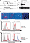
CLEC-2–deficient platelets do not interact with PDPN. (A) Gene targeting strategy for Clec-2. The deleted sequence in exon 1 encodes the entire intracellular domain of the CLEC-2 receptor, including the YxxL motif required to activate SYK, as well as the transcriptional and translational start sites. (B) Clec-2−/− platelets lack CLEC-2 protein. Immunoblotting was used to measure CLEC-2 and actin proteins in cell lysate derived from brain (left lane) and platelet-rich plasma (right lanes). CLEC-2 levels in Clec-2+/− platelets were reduced by approximately 50% compared with those in Clec-2+/+ platelets (second and third lanes from left, lysate from 0.75 mL of blood loaded). BecauseClec-2−/− mice die in the perinatal period, CLEC-2 levels were measured in platelets derived from lethally irradiated wild-type mice that had been reconstituted with Clec-2+/+ or Clec-2−/− fetal liver cells (right 4 lanes, lysate from 0.1 mL of blood loaded in each lane). (C) In situ hybridization of E14.5 mouse embryos for Clec-2. Clec-2 expression is indicated in red; DAPI nuclear staining is shown in blue. (D) PDPN-Fc proteins bind Clec-2+/+ but not Clec-2−/− platelets. (E) PDPN-Fc proteins activate Clec-2+/+ but not Clec-2−/− platelets. Platelet activation was measured by the binding of allophycocyanin-conjugated fibrinogen. Platelet activation by the glycoprotein VI receptor agonist convulxin is shown as a positive control.
Figure 3
Figure 3
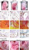
CLEC-2 is required in blood cells for normal lymphatic vascular development. (A-L) Clec-2−/− animals exhibit blood-lymphatic vascular mixing phenotypes. (A-F) Clec-2−/− embryos exhibit blood-filled lymphatics like those of Slp-76−/− animals during mid-gestation. Whole embryo images show blood-filled cutaneous lymphatics and edema (above). Transverse sections show anti-LYVE-1 immunostaining (brown) of lymph sacs adjacent to the cardinal vein (below). (G-L) Clec-2−/− neonates exhibited blood-filled mesenteric vessels (G,I,K) and edema of the intestine wall (H,J,L) like those of Slp-76−/− animals. (M-O) Blood-filled mesenteric lymphatic vessels arise in lethally irradiated wild-type mice after reconstitution with Clec-2−/− (N-O) but not wild-type (M) donor hematopoietic cells. A indicates artery; V, vein; and L, lymphatic.
Figure 4
Figure 4
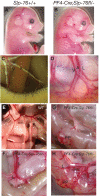
SLP-76 is required in platelets for normal lymphatic vascular development. (A-B) PF4-Cre;Slp-76fl/ embryos exhibit blood-filled cutaneous lymphatics. (C-D) PF4-Cre;Slp-76fl/ neonates exhibit blood-filled mesenteric and intestinal lymphatic vessels. A indicates artery; V, vein; and L, lymphatic. (E-H) Twelve-week-old PF4-Cre;Slp-76fl/ animals exhibit vascular mixing phenotypes that range in severity from blood-filled Peyer patches (PP, left) to disruption of the normal vascular pattern of the intestine wall and blood-filled mesenteric lymphatic vessels (L, right).
Figure 5
Figure 5
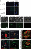
Platelets are activated by and form aggregates on LECs in a SLP-76–dependent manner in vitro and in vivo. (A) Wild-type platelets bind and are activated on the surface of LECs but not BECs. Slp-76−/− platelets adhere to LECs but do not form aggregates or express P-selectin. CD41 staining identifies platelets, and P-selectin expression identifies activated platelets. (B) Flow of heparinized wild-type blood at a shear level of 8 dynes/cm2 results in platelet aggregate formation on LECs but not on BECs (left 2 panels). PDPN-deficient platelets adhere and form aggregates on LECs, but SLP-76–deficient platelets fail to form aggregates (right 2 panels). Platelets are visualized with Alexa Fluor 488–conjugated anti-CD41 antibody after 5 minutes of flow. (C) Identification of platelet aggregates on LYVE-1+ LECs in Slp-76+/+ but not Slp-76−/− developing embryos. Immunostaining of E11.5 transverse embryo sections with antibodies against platelets (red) and the LEC LYVE-1 protein (green) are shown. Arrows indicate platelet aggregates on the endothelial cell surface. CV indicates cardinal vein; and LS, lymph sac. (D-E) Injection of wild-type platelets into Slp-76−/− animals results in the formation of platelet aggregates on the surface of LECs. Wild-type platelets were injected into the vitelline vein of E18 Slp-76−/− embryos (D) or into the retroorbital sinus of P21 Slp-76−/− animals (E). Platelet aggregates (red) and lymphatic endothelium (anti–LYVE-1, green) were detected by immunostaining of tissues harvested 12 hours after injection.
Figure 6
Figure 6
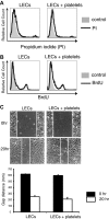
Platelets do not affect LEC viability, proliferation, or migration in vitro.(A) Overnight culture of platelets with LECs does not affect endothelial cell viability measured by nuclear uptake of propidium iodide. Control indicates cells not treated with propidium iodide. (B) Overnight culture of platelets with LECs does not affect endothelial cell proliferation measured by uptake of BrdU after a 2-hour pulse. Control indicates cells not treated with BrdU. (C) Culture of platelets with LECs does not alter migration in a scratch assay. N = 3 for each group. Error bars indicate SD.
Figure 7
Figure 7
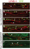
Blood cells do not contribute to lymphatic endothelium at the site of vascular mixing in mice lacking PDPN, CLEC-2, or SLP-76. Vav-Cre–activated YFP expression was used to detect hematopoietic contribution to lymphatic endothelium in the intestine, the site of greatest vascular mixing in mice lacking PDPN, CLEC-2, or SLP-76. (A-D) Confocal microscopy of sections of Vav-Cre;R26RYFP neonates immunostained for the lymphatic endothelial surface protein LYVE-1 (red) and YFP (green) fails to show YFP+LYVE-1+ endothelial cells. Arrows indicate LYVE-1+ endothelial cells. Arrowheads indicate YFP+ hematopoietic cells that are frequently adjacent to but not incorporated within LYVE-1+ endothelium. (E-F) Analysis of Tie2-Cre;R26RYFP neonates shows numerous true LYVE-1+YFP+ endothelial cells (arrows).

Comment in

  • Inside bloody lymphatics.
    D'Amico G, Alitalo K. D'Amico G, et al. Blood. 2010 Jul 29;116(4):512-3. doi: 10.1182/blood-2010-04-278549. Blood. 2010. PMID: 20671134 No abstract available.

References

    1. Shalaby F, Rossant J, Yamaguchi TP, et al. Failure of blood-island formation and vasculogenesis in Flk-1-deficient mice. Nature. 1995;376(6535):62–66. - PubMed
    1. Stainier DY, Weinstein BM, Detrich HW, III, Zon LI, Fishman MC. Cloche, an early acting zebrafish gene, is required by both the endothelial and hematopoietic lineages. Development. 1995;121(10):3141–3150. - PubMed
    1. Chen MJ, Yokomizo T, Zeigler BM, Dzierzak E, Speck NA. Runx1 is required for the endothelial to haematopoietic cell transition but not thereafter. Nature. 2009;457(7231):887–891. - PMC - PubMed
    1. Rafii S, Lyden D. Therapeutic stem and progenitor cell transplantation for organ vascularization and regeneration. Nat Med. 2003;9(6):702–712. - PubMed
    1. Purhonen S, Palm J, Rossi D, et al. Bone marrow-derived circulating endothelial precursors do not contribute to vascular endothelium and are not needed for tumor growth. Proc Natl Acad Sci U S A. 2008;105(18):6620–6625. - PMC - PubMed

Publication types

MeSH terms