. 2011 Jan;21(1):22-34.
doi: 10.1093/cercor/bhq051. Epub 2010 Apr 2.

What goes down must come up: role of the posteromedial cortices in encoding and retrieval

Affiliations

What goes down must come up: role of the posteromedial cortices in encoding and retrieval

P Vannini et al. Cereb Cortex. 2011 Jan.

Abstract

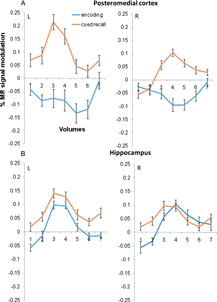
The hypothesis that the neural network supporting successful episodic memory retrieval overlaps with the regions involved in episodic encoding has garnered much interest; however, the role of the posteromedial regions remains to be fully elucidated. Functional magnetic resonance imaging (fMRI) studies during successful encoding typically demonstrate deactivation of posteromedial cortices, whereas successful retrieval of previously encoded information has been associated with activation of these regions. Here, we performed an event-related fMRI experiment during an associative face-name encoding and retrieval task to investigate the topography and functional relationship of the brain regions involved in successful memory processes. A conjunction analysis of novel encoding and subsequent successful retrieval of names revealed an anatomical overlap in bilateral posteromedial cortices. In this region, a significant negative correlation was found: Greater deactivation during encoding was related to greater activation during successful retrieval. In contrast, the hippocampus and prefrontal cortex demonstrated positive activation during both encoding and retrieval. Our results provide further evidence that posteromedial regions constitute critical nodes in the large-scale cortical network subserving episodic memory. These results are discussed in relation to the default mode hypothesis, the involvement of posteromedial cortices in successful memory formation and retention, as well as potential implications for aging and neurodegenerative disease.


Figures

Figure 1.
Figure 1.
fMRI paradigm. (A) The paradigm consisted of 4 encoding runs alternating with 4 retrieval runs. In each encoding run, 20 face–name pairs were presented to the subjects 3 times (EN1, EN2, and EN3). Each encoding run was immediately followed by a retrieval run, consisting of a cued recall (CR) task followed by a forced-choice recognition (FCR) task for each of the 20 face–name pairs in the preceding encoding run. (B) The experimental setup of the first encoding run followed by the first retrieval run. In encoding run 1, the first group of 4 face–name pairs (5 groups of 4 face–name pairs comprised the total of 20 stimuli for each encoding run) is displayed (EN1a) and how the stimuli were repeated over the run (EN2a and EN3a), each time presented in a pseudorandomised order within the group (see numbers on the top of the figure). For each face–name pair, the subjects were asked to press a button indicating a purely subjective decision about whether the name was a good “fit” for the face or not. The figure also displays the same 4 faces (RETa) in the first retrieval run. In the CR task, the subjects had to respond with a button press whether he/she “remembered” the name associated with the face or had “forgotten” it. During the FCR task, the subjects were instructed to indicate the correct name associated with that face by pressing 1 of the 2 buttons (correct name was presented in counterbalanced order across all runs).
Figure 2.
Figure 2.
Brain regions involved in the face–name association task. Statistical parametric maps (random effects) from the one sample t-test demonstrating the deactivation in encoding (A) and activation during cued recall (B) as well as the conjunction (C) of these 2 statistical maps, demonstrating a significant overlap in the precuneus and posterior cingulate regions. In the lower row, the statistical parametric maps from the one sample t-test illustrates the activation in encoding (D) and during cued recall (E) as well as the conjunction (F) between these 2, demonstrating significant overlap in activation in the hippocampus. The maps are threshold at P < 0.001, minimal extent threshold 5 adjacent voxels and superimposed on a single-subject high-resolution T1 structural images, AC on Talairach sagittal planes, x = 12 and DF on Talairach coronal planes, y = 15. Lighter color scale indicates more significant activation (orange/red) or deactivation (blue).
Figure 3.
Figure 3.
MR signal time courses from overlapping brain regions during successful encoding and retrieval. Extracted % MR signal time courses (time repetition = 2 s) during successful encoding (blue) and successful cued recall (orange) from region of interest in posteromedial cortex (A) left precuneus (x = −6, y = −65, z = 36) and right retrosplenial cortex (x = 21, y = −54, z = 28) and hippocampus (B) left (x = −27, y = −15, z = 12) and right (x = 24, y = −24, z = −6). Error bars represent standard error.
Figure 4.
Figure 4.
Relationship between the beta weights during encoding and retrieval in the posteromedial cortices. Extracted beta weights during successful encoding (blue) and successful cued recall (orange) from the right retrosplenial VOI (x = 21, y = −54, z = 28). Error bars represent standard error. A negative correlation was found between the 2 variables (r = −0.44, P = 0.05).
Figure 5.
Figure 5.
(A) One sample t-test of Remember Hit > Fix (left) and Forgotten Hit > Fix (right) contrasts. The latter was made in order to investigate which areas were activated when the subjects said that they had forgotten the name during the cued recall test but choose the right name during the forced choice recognition test. The activation maps are projected on an inflated (200 iterations) template brain surface. A surface based smoothing (10 mm) was applied. Statistical values are displayed on the surface in color scale, thresholded at p = 0.001 with an extent threshold of 5 voxels. (B) Anatomical overlap in fMRI activity from exploratory analysis. Conjunction map of (A) RHITenc > FORGET AND RHITret > FORGET demonstrating overlap of activation in hippocampus and (B) FORGET > RHITenc AND RHITret > FORGET showing overlap of deactivation and activation in posteromedial cortex. The maps are threshold at P < 0.05, minimal extent threshold 5 adjacent voxels and superimposed on a single-subject high-resolution T1 images. Lightness in center of colored area indicates more significant activity. Extracted beta weights during successful encoding (light blue) and FORGET (i.e., weighted mean for FHIT and FMISS) (dark blue) and successful cued recall (light orange) and FORGET (dark orange) from the VOIs in the left precuneus (global maximum found in Tal coordinates [−15 −68 42]) and right hippocampus (21 −9 −15).

References

    1. Addis DR, McIntosh AR, Moscovitch M, Crawley AP, McAndrews MP. Characterizing spatial and temporal features of autobiographical memory retrieval networks: a partial least squares approach. Neuroimage. 2004;23:1460–1471. - PubMed
    1. Addis DR, Wong AT, Schacter DL. Remembering the past and imagining the future: common and distinct neural substrates during event construction and elaboration. Neuropsychologia. 2007;45:1363–1377. - PMC - PubMed
    1. Alvarez P, Squire LR. Memory consolidation and the medial temporal lobe: a simple network model. Proc Natl Acad Sci U S A. 1994;91:7041–7045. - PMC - PubMed
    1. Brett M, Anton J, Valabregue R, Poline J-B. Region of interest analysis using an SPM toolbox. 2002 At the 8th International Conference on Functional Mapping of the Human Brain; Sendai, Japan.
    1. Buckner RL, Andrews-Hanna JR, Schacter DL. The brain’s default network: anatomy, function, and relevance to disease. Ann N Y Acad Sci. 2008;1124:1–38. - PubMed

Publication types