. 2010 Jul;334(1):33-43.
doi: 10.1124/jpet.109.163840. Epub 2010 Apr 2.

Human recombinant vascular endothelial growth factor reduces necrosis and enhances hepatocyte regeneration in a mouse model of acetaminophen toxicity

Affiliations

Human recombinant vascular endothelial growth factor reduces necrosis and enhances hepatocyte regeneration in a mouse model of acetaminophen toxicity

Brian C Donahower et al. J Pharmacol Exp Ther. 2010 Jul.

Abstract

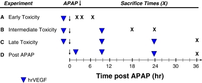
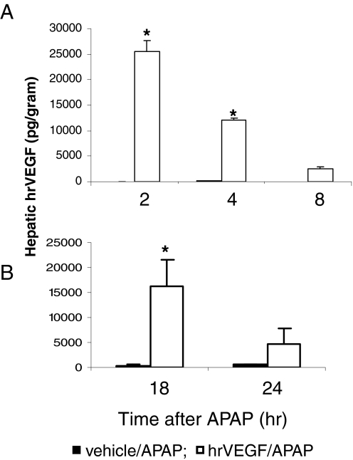
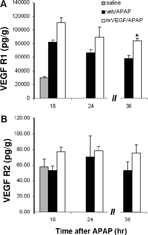
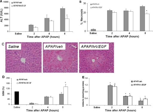
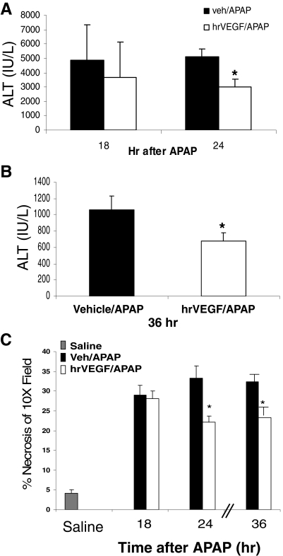
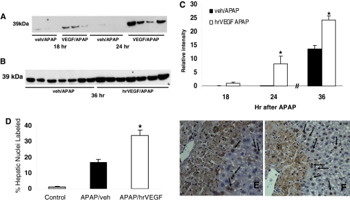
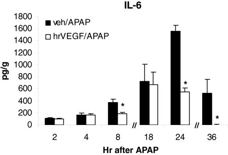
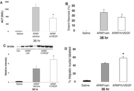
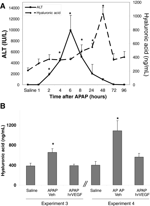
We reported previously that vascular endothelial growth factor (VEGF) was increased in acetaminophen (APAP) toxicity in mice and treatment with a VEGF receptor inhibitor reduced hepatocyte regeneration. The effect of human recombinant VEGF (hrVEGF) on APAP toxicity in the mouse was examined. In early toxicity studies, B6C3F1 mice received hrVEGF (50 microg s.c.) or vehicle 30 min before receiving APAP (200 mg/kg i.p.) and were sacrificed at 2, 4, and 8 h. Toxicity was comparable at 2 and 4 h, but reduced in the APAP/hrVEGF mice at 8 h (p < 0.05) compared with the APAP/vehicle mice. Hepatic glutathione (GSH) and APAP protein adduct levels were comparable between the two groups of mice, with the exception that GSH was higher at 8 h in the hrVEGF-treated mice. Subsequently, mice received two doses (before and 10 h) or three doses (before and 10 and 24 h) of hrVEGF; alanine aminotransferase values and necrosis were reduced at 24 and 36 h, respectively, in the APAP/hrVEGF mice (p < 0.05) compared with the APAP/vehicle mice. Proliferating cell nuclear antigen expression was enhanced, and interleukin-6 expression was reduced in the mice that received hrVEGF (p < 0.05) compared with the APAP/vehicle mice. In addition, treatment with hrVEGF lowered plasma hyaluronic acid levels and neutrophil counts at 36 h. Cumulatively, the data show that treatment with hrVEGF reduced toxicity and increased hepatocyte regeneration in APAP toxicity in the mouse. Attenuation of sinusoidal cell endothelial dysfunction and changes in neutrophil dynamics may be operant mechanisms in the hepatoprotection mediated by hrVEGF in APAP toxicity.


Figures

Fig. 1.
Fig. 1.
Summary of study designs. Four experimental designs were used in the study. A–C, hrVEGF was administered as 50 μg s.c. (denoted by ▼), and mice were sacrificed at various times (denoted by X) to characterize the effect of hrVEGF on the early (A), intermediate (B), and late (C) stages of toxicity. D, a rescue design study was performed in which hrVEGF treatment was initiated 2 h after APAP administration.
Fig. 2.
Fig. 2.
Demonstration of hrVEGF in mouse liver after treatment with hrVEGF. A, mice were treated with APAP (200 mg/kg i.p.) plus vehicle (PBS) or APAP plus hrVEGF (50 μg, s.c.) and sacrificed at the indicated times. hVEGF levels were significantly elevated at 2 and 4 h in mice treated with hrVEGF. B, mice were treated with APAP and received two doses of hrVEGF or vehicle and were sacrificed at the indicated times. hVEGF levels were significantly elevated at 18 h in mice treated with hrVEGF. hVEGF levels in PBS-treated mice were 0.0 pg/g in both experiments (data not shown). *, significant difference from APAP/veh groups, p < 0.05.
Fig. 3.
Fig. 3.
Effect of hrVEGF on VEGFR1 (A) and VEGFR2 (B) expression. VEGFR1 and VEGFR2 levels were examined in the mice from the early (not shown), intermediate, and late experiments. Mean (± S.E.) hepatic VEGFR1 and VEGFR2 levels were increased in the hrVEGF-treated mice compared with the mice that received vehicle/APAP. This finding was significant for VEGFR1 in the 36-h experiment. *, p < 0.05.
Fig. 4.
Fig. 4.
Effect of hrVEGF on ALT, histology, and metabolism. Mice were treated with APAP/veh or APAP/hrVEGF and sacrificed at the indicated times. A, at 8 h, mice receiving hrVEGF plus APAP had significantly reduced ALT levels compared with the mice that received vehicle plus APAP. *, p < 0.05, significant difference from APAP/veh group. B, histological examination of H&E-stained slides revealed decreased necrosis in the hrVEGF-treated mice at 8 h. C, histology of saline-, APAP/veh-, and APAP/hrVEGF-treated mice. D, at 2 and 4 h, APAP/hrVEGF mice and APAP/veh mice had comparable reductions in GSH levels. At 8 h, hrVEGF-treated mice had significantly higher GSH levels than the APAP/veh mice. *, p < 0.05, significant difference from APAP/veh group. E, APAP protein adducts in liver homogenates of mice treated with APAP/veh or APAP/hrVEGF. Adducts were increased at 2, 4, and 8 h in all treatment groups. No differences were present between the APAP/veh and the APAP/hrVEGF groups at any time point.
Fig. 5.
Fig. 5.
Effect of hrVEGF on ALT levels in the intermediate and late stages of toxicity. Mice were treated with APAP/veh or APAP/hrVEGF and sacrificed at the indicated times. A, at 18 and 24 h, mice receiving hrVEGF had reduced ALT levels compared with the APAP/veh mice. Mean (± S.E.) ALT values in PBS mice were 28.9 ± 5.3 IU/liter (data not shown). B, mice treated with hrVEGF had significantly lower ALT levels than vehicle-treated mice at 36 h. Mean ALT (± S.E.) values in control mice were 16.2 ± 2.1 IU/liter (data not shown). *, p < 0.05, significant difference from veh/APAP group. C, necrosis scores of mice sacrificed at 18, 24, and 36 h. Necrosis was reduced (*, p < 0.05) in the hrVEGF-treated mice at 24 and 36 h compared with the APAP/veh-treated mice.
Fig. 6.
Fig. 6.
Effect of hrVEGF on PCNA expression. A, mice were treated with APAP/veh or APAP/hrVEGF and sacrificed at 18 or 24 h. hrVEGF treatment increased PCNA expression by Western blot at 18 and at 24 h compared with APAP/veh-treated mice. B, mice were treated with APAP/veh or APAP/hrVEGF and sacrificed at 36 h. Mice treated with hrVEGF had increased expression of PCNA by Western blot compared with vehicle-treated mice. C, densitometric analysis indicated a significant difference in PCNA expression, represented as mean (±S.E.), at 24 and 36 h in the hrVEGF-treated mice (p < 0.05) compared with the APAP/veh mice at the same time points. D, image analysis of PCNA-stained slides from the 36-h experiment indicated increased PCNA labeling in the hepatic nuclei of the mice treated with hrVEGF compared with the vehicle-treated mice. *, p < 0.05. E, representative mouse liver section of PCNA staining in an APAP/veh-treated mouse. F, representative mouse liver section of PCNA staining from an APAP/hrVEGF-treated mouse.
Fig. 7.
Fig. 7.
Effect of hrVEGF on hepatic IL-6 expression. IL-6 levels were examined in the mice from the early, intermediate, and late experiments. Mean (± S.E.) hepatic IL-6 levels were reduced in mice that received hrVEGF compared with vehicle-treated mice. *, p < 0.05.
Fig. 8.
Fig. 8.
Effect of treatment with hrVEGF on ALT levels, necrosis, and PCNA expression at 36 h. Mice were treated with APAP followed by hrVEGF or vehicle at 2, 10, or 24 h and sacrificed at 36 h. A, at 36 h, mice receiving hrVEGF had reduced ALT levels compared with the APAP/veh mice. B, mice treated with hrVEGF had significantly reduced necrosis scores compared with the vehicle-treated mice. C, PCNA expression by immunoblot. Mice treated with hrVEGF had increased expression of PCNA by immunoblot analysis. Lanes signify representative mice, and densitometry represents summary of all mice (n = 12 per treatment group). D, summary of image analysis for PCNA-labeled nuclei. *, p < 0.05 for all comparisons of APAP/veh versus APAP/hrVEGF.
Fig. 9.
Fig. 9.
Effect of hrVEGF on plasma levels of HA. A, mice were treated with APAP (200 mg/kg i.p.) and sacrificed at the indicated times. Plasma HA levels were significantly increased at 4, 8, 24, and 48 h (p < 0.05), and the onset of plasma HA elevation occurred before the ALT response. B, plasma HA levels were measured in mice that received hrVEGF before APAP and 2 h after APAP. Additional doses of hrVEGF were administered at 10 and 24 h, and the mice were sacrificed at 36 h. Plasma HA levels were reduced at 36 h in both groups of mice treated with hrVEGF. *, p < 0.05.

References

    1. Bajt ML, Knight TR, Farhood A, Jaeschke H. (2003) Scavenging peroxynitrite with glutathione promotes regeneration and enhances survival during acetaminophen-induced liver injury in mice. J Pharmacol Exp Ther 307:67–73 - PubMed
    1. Banai S, Jaklitsch MT, Shou M, Lazarous DF, Scheinowitz M, Biro S, Epstein SE, Unger EF. (1994) Angiogenic-induced enhancement of collateral blood flow to ischemic myocardium by vascular endothelial growth factor in dogs. Circulation 89:2183–2189 - PubMed
    1. Bourdi M, Masubuchi Y, Reilly TP, Amouzadeh HR, Martin JL, George JW, Shah AG, Pohl LR. (2002) Protection against acetaminophen-induced liver injury and lethality by interleukin 10: role of inducible nitric oxide synthase. Hepatology 35:289–298 - PubMed
    1. Conn G, Soderman DD, Schaeffer MT, Wile M, Hatcher VB, Thomas KA. (1990) Purification of a glycoprotein vascular endothelial cell mitogen from a rat glioma-derived cell line. Proc Natl Acad Sci USA 87:1323–1327 - PMC - PubMed
    1. Cressman DE, Greenbaum LE, DeAngelis RA, Ciliberto G, Furth EE, Poli V, Taub R. (1996) Liver failure and defective hepatocyte regeneration in interleukin-6-deficient mice. Science 274:1379–1383 - PubMed

Publication types

MeSH terms