. 2010 Jun;298(6):H1738-47.
doi: 10.1152/ajpheart.00688.2009. Epub 2010 Apr 2.

Effects of cardiac-restricted overexpression of the A(2A) adenosine receptor on adriamycin-induced cardiotoxicity

Affiliations

Effects of cardiac-restricted overexpression of the A(2A) adenosine receptor on adriamycin-induced cardiotoxicity

Eman A Hamad et al. Am J Physiol Heart Circ Physiol. 2010 Jun.

Abstract

Activation of the A(2A) adenosine receptor (A(2A)R) has been shown to be cardioprotective. We hypothesized that A(2A)R overexpression could protect the heart from adriamycin-induced cardiomyopathy. Transgenic (TG) mice overexpressing the A(2A)R and wild-type mice (WT) were injected with adriamycin (5 mg.kg(-1).wk(-1) ip, 4 wk). All WT mice survived adriamycin treatment while A(2A)R TG mice suffered 100% mortality at 4 wk. Telemetry showed progressive prolongation of the QT interval, bradyarrhythmias, heart block, and sudden death in adriamycin-treated A(2A)R TG but not WT mice. Both WT and A(2A)R TG demonstrated similar decreases in heart function at 3 wk after treatment. Adriamycin significantly increased end-diastolic intracellular Ca(2+) concentration in A(2A)R TG but not in WT myocytes (P < 0.05). Compared with WT myocytes, action potential duration increased dramatically in A(2A)R TG myocytes (P < 0.05) after adriamycin treatment. Expression of connexin 43 was decreased in adriamycin treated A(2A)R TG but not WT mice. In sharp contrast, A(2A)R overexpression induced after the completion of adriamycin treatment resulted in no deaths and enhanced cardiac performance compared with WT adriamycin-treated mice. Our results indicate that the timing of A(2A)R activation is critical in terms of exacerbating or protecting adriamycin-induced cardiotoxicity. Our data have direct relevance on the clinical use of adenosine agonists or antagonists in the treatment of patients undergoing adriamycin therapy.


Figures
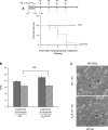
Fig. 1.
Fig. 1.
A: survival after adriamycin (Ad) treatment. All wild-type (WT) mice survived adriamycin treatment, while adenosine A2A receptor (A2AR) transgenic (TG) mice had 100% mortality by the end of 4 wk (P < 0.05, log-rank test, n = 10). DOX, doxycyclin. B: evaluation of cardiac function after third adriamycin treatment in A2AR TG and WT mice. Adriamycin reduced cardiac function in both WT and A2ATG mice. Percent fractional shortening (FS) of indicated mouse groups is shown. *P < 0.01 WT baseline vs. WT adriamycin. +P < 0.001 A2AR TG baseline vs. A2AR TG adriamycin. NS, not significant WT adriamycin vs. A2AR TG adriamycin (see Table 1 for details). C: electron microscopic images of myocardial sections of WT (n = 2) and A2ATG (n = 2) mouse hearts after third adriamycin injection. m, Mitochondrion. Bar = 500 nM.
Fig. 2.
Fig. 2.
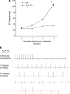
Telemetric tracings from A2AR TG and WT mice treated with adriamycin. A: telemetric data from A2AR TG and WT mice (n = 3 for each group). Progressive prolongation of QT segment (ms) with each injection in A2AR TG (P < 0.001) is shown. WT mice had some prolongation in QT interval after the third injection, but they had no arrhythmias. *P < 0.05 (n = 3 mice for each group, male 10- to 12-wk-old mice). B: development of runs of ventricular tachycardia, bradyarrhythmias, and first- and second-degree heart block.
Fig. 3.
Fig. 3.
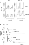
Intracellular Ca2+ concentration ([Ca2+]i) transients and action potential durations. After adult myocytes from WT and A2ATG were isolated, 5 μM adriamycin was added to the culture medium and measurements were made 18 h after exposure. A: representative tracing of myocyte calcium transients in A2AR TG and WT mice with and without adriamycin. B: representative tracing of action potential duration measurements in A2AR TG and WT mice with and without adriamycin.
Fig. 4.
Fig. 4.
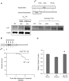
Connexin 43 (Cx43) staining patterns and expression in A2AR TG and WT mice treated with adriamycin. A: after the third dose of adriamycin, ventricular myocardium from A2AR TG and WT mice was immunostained with Cx43 and β-catenin (n = 3 for each group). B: ventricular extracts were prepared from hearts not exposed to adriamycin (left) and from hearts after 3 injections of adriamycin (right). Ten- to 12-wk-old male WT hearts (n = 4) and A2A TG hearts (n = 4–6) were probed for Cx43 and dephosphorylated (De-P Cx43, 13-8300). Cx43 signals were normalized to GAPDH. Values are means ± SE. *P < 0.05 vs. WT.
Fig. 5.
Fig. 5.
Effect of A2AR expression after cessation of adriamycin treatment. A: schematic diagram showing DOX inhibition and induction of A2AR in 8-wk-old TG mice. Mice were harvested at indicated times after DOX removal. Total ventricular protein extracts were immunoblotted with anti-A2AR antibody. B: schematic diagram showing induction of A2AR expression after cessation of adriamycin administration. Survival curve showed that control mice treated with adriamycin (n = 24) had over 30% mortality, while all A2AR TG mice (n = 7) survived. C: relative cardiac function in A2AR TG and control mice 8 wk after adriamycin administration. As detailed in materials and methods, this echocardiographic experiment was performed using the ACUSON Sequoia C256 system and Avertin anesthesia. Exact %FS: WT no adriamycin (53.0 ± 1.4, n = 8), WT post-adriamycin (45.2.0 ± 1.9, n = 12); A2AR TG post-adriamycin (52.7 ± 1.3, n = 6). *P < 0.05 vs. WT post-adriamycin.

References

    1. Akar FG, Spragg DD, Tunin RS, Kass DA, Tomaselli GF. Mechanisms underlying conduction slowing and arrhythmogenesis in nonischemic dilated cardiomyopathy. Circ Res 95: 717–725, 2004 - PubMed
    1. Beardslee MA, Lerner DL, Tadros PN, Laing JG, Beyer EC, Yamada KA, Kleber AG, Schuessler RB, Saffitz JE. Dephosphorylation and intracellular redistribution of ventricular connexin43 during electrical uncoupling induced by ischemia. Circ Res 87: 656–662, 2000 - PubMed
    1. Bristow MR, Minobe WA, Billingham ME, Marmor JB, Johnson GA, Ishimoto BM, Sageman WS, Daniels JR. Anthracycline-associated cardiac and renal damage in rabbits. Evidence for mediation by vasoactive substances. Lab Invest 45: 157–168, 1981 - PubMed
    1. Buja LM, Ferrans VJ, Mayer RJ, Roberts WC, Henderson E. Cardiac ultrastnictural changes induced by daunorubicin therapy. Cancer 32: 771–778, 1973 - PubMed
    1. Chan TO, Funakoshi H, Song J, Zhang XQ, Wang J, Chung PH, Degeorge BR, Li X, Zhang J, Herrmann DE, Diamond M, Hamad E, Houser SR, Koch WJ, Cheung JY, Feldman AM. Cardiac-restricted overexpression of the A2A-adenosine receptor in FVB mice transiently increases contractile performance and rescues the heart failure phenotype in mice overexpressing the A1-adenosine receptor. Clin Transl Sci 1: 126–133, 2008 - PMC - PubMed

Publication types

MeSH terms