. 2010 May;176(5):2467-76.
doi: 10.2353/ajpath.2010.090519. Epub 2010 Apr 2.

Impaired gastric gland differentiation in Peutz-Jeghers syndrome

Affiliations

Impaired gastric gland differentiation in Peutz-Jeghers syndrome

Lina Udd et al. Am J Pathol. 2010 May.

Abstract

Gastrointestinal hamartomatous polyps in the Peutz-Jeghers cancer predisposition syndrome and its mouse model (Lkb1(+/-)) are presumed to contain all cell types native to the site of their occurrence. This study aimed to explore the pathogenesis of Peutz-Jeghers syndrome polyposis by characterizing cell types and differentiation of the epithelium of gastric polyps and predisposed mucosa. Both antral and fundic polyps were characterized by a deficit of pepsinogen C-expressing differentiated gland cells (antral gland, mucopeptic, and chief cells); in large fundic polyps, parietal cells were also absent. Gland cell loss was associated with an increase in precursor neck cells, an expansion of the proliferative zone, and an increase in smooth muscle alpha-actin expressing myofibroblasts in the polyp stroma. Lack of pepsinogen C-positive gland cells identified incipient polyps, and even the unaffected mucosa of young predisposed mice displayed an increase in pepsinogen C negative glands (25%; P = 0045). In addition, in small intestinal polyps, gland cell differentiation was defective, with the absence of Paneth cells. There were no signs of metaplastic differentiation in any of the tissues studied, and both the gastric and small intestinal defects were seen in Lkb1(+/-) mice, as well as polyps from patients with Peutz-Jeghers syndrome. These results identify impaired epithelial differentiation as the earliest pathological sign likely to contribute to tumorigenesis in individuals with inherited Lkb1 mutations.


Figures

Figure 1
Figure 1
Abnormally proportioned gastric units and lack of terminal gland cell differentiation in large gastric Lkb1+/− polyps with characteristic Peutz-Jeghers features. A: Comparison of proliferating (Ki-67), foveolar (Alcian blue-Periodic acid-Schiff-; AB-PAS) and glandular cells (Griffonia Simplicifolia-lectin; GS-lectin) in antral mucosa of wild-type and Lkb1+/− mice, and in an antral polyp demonstrating a widening of the proliferating, foveolar (f), and glandular (g) zones in an antral Lkb1+/− polyp, at ×200 magnification. Scale bars = 100 μm. B: Staining of an antral polyp with desmin, GS-lectin, and AB-PAS demonstrates that GS-lectin positive gland-type cells are more abundant in the vicinity of desmin staining smooth muscle bundles (arrowheads). Micrographs recorded at ×100 magnification. Size bars = 200 μm. C: A fundic polyp demonstrating lack of pepsinogen C expressing zymogenic/chief cells and parietal cells (Hydrogen-potassium adenosine-triphosphatase; H+/K+-ATPase), whereas GS-lectin positive mucous neck cells (n) and foveolar cells (AB-PAS, arrowhead) are abundant. Inset in GS-lectin panel demonstrates that GS-lectin positive mucous neck cells (n) cover the gland base adjacent to the smooth muscle stalk (m), whereas in normal glands gland the base contains chief cells (c). The surface of this polyp had suffered an erosion (asterisks).
Figure 2
Figure 2
Aberrant epithelium and stroma in incipient Lkb1+/− polyps. A: Staining of proliferating (Ki-67) and pyloric gland cells (pepsinogen C) in antral mucosa of wild-type and Lkb1+/− mice, and in an incipient polyp (bracket). All micrographs at ×100 magnification. Scale bars = 200 μm. The normal gastric folds of wild-type antral mucosa cause glancing of the section (*) at the proliferative zone (Ki-67) without disrupting the pepsinogen C staining pattern (arrowhead). B: Decreased pepsinogen C expression in an incipient antral Lkb1+/− polyp (bracket) is associated with accumulation of neck cells (GS-lectin, arrow), and an increase in smooth muscle α-actin (SMA) expression (arrowheads; ×100 magnification. Scale bar = 200 μm. C: Pepsinogen C staining of antral mucosa in 4.5-month-old Lkb1+/− mice reveals an increase in pepsinogen C deficient glands (pepsinogen altered pyloric glands, PAPG, marked with asterisks in micrograph) compared with the wild-type. The frequencies of PAPG (PAPG %) are shown in the bar chart, n = number of glands (number of mice in parentheses). D: Triple staining with SMA (black chromogen), pepsinogen C (red chromogen), and Ki-67 (brown chromogen) of antral mucosa from a 4.5-month-old mouse demonstrates pepsinogen C deficient glands (left panel, asterisks; ×200 magnification) and Ki-67 positive proliferating cells (arrowhead) at the base of a pepsinogen C negative gland in the right panel (×630 magnification).
Figure 3
Figure 3
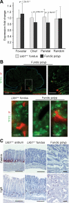
Alterations in the expression of cell-type specific gene sets in Lkb1+/− tissues, and the exclusion of spasmolytic polypeptide-expressing metaplasia in Lkb1+/− fundic polyps. A: Average fold change ± SD of gene expression in indicated cell-type specific gene sets including a random set for comparison (please see Materials and Methods) in Lkb1+/− polyps (black) and unaffected fundic mucosa (gray) compared with wild-type mucosa. P values indicate the statistical significance of the difference between the wild-type and the corresponding value, as given by the Student’s t-test. B: TFF2 and IF staining of Lkb1+/− fundic polyps and wild-type mucosa, demonstrating absence of IF in the polyp (upper row) at ×100, and a magnification of the area within the white box in the upper right panel. Scale bars = 200 μm. In the lower panels, micrographs at ×630 magnification (Scale bars = 32 μm) show fundic Lkb1+/− mucosa (left panel) and only TFF2-staining gland cells in the polyp proper (middle panel) as well as a gland with a single IF-positive cell from the border of a polyp base (right panel). C: Gastrin and chromogranin A (CgA) staining of Lkb1+/− antrum, fundus and a fundic polyp provide no evidence for antralization (antrum and fundus at ×200 magnification; polyp at ×100 magnification; Scale bars = 100 μm).
Figure 4
Figure 4
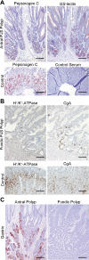
Impaired gland cell differentiation in human gastric Peutz-Jeghers polyps. A: Pepsinogen C or GS-lectin staining of sections from the base of an antral Peutz-Jeghers patient polyp or control gastric mucosa from a patient biopsy specimen (all micrographs at ×100 magnification; Scale bar = 200 μm). B: Staining for H+/K+-ATPase demonstrates deficiency in parietal cells in the polyp and staining for chromogranin A (CgA) shows presence of enteroendocrine cells in the base of a fundic Peutz-Jeghers patient polyp (at ×100 magnification; Scale bar = 200 μm). C: Comparison of gastrin positive cells in an antral and fundic polyp indicates presence according to the native site of occurrence (also at ×100 magnification; Scale bar = 200 μm).
Figure 5
Figure 5
Correct specification into gastric versus intestinal epithelium in Peutz-Jeghers polyps and impaired Paneth cell differentiation in intestinal polyps. A: AB-PAS and TFF2 stainings of Lkb1+/− polyps from jejunum and antrum, indicating intestinal (acid) type mucin in cells with goblet morphology in the jejunal polyp consistent with normal jejunal staining pattern, and gastric (neutral) mucin on the surface of the antral polyp, as in normal gastric mucosa (micrograph at ×400; Scale bar = 50 μm). TFF2 is expressed in an antral polyp, and in the jejunal polyp there is no ectopic expression (middle panels, overlaid to the right with guides highlighting the basal surface of the epithelium in magenta, and the apical surface in blue), at ×200 magnification; Scale bar = 100 μm. B: Staining of intestinal transcription factor Cdx2 in fundic and small intestinal Lkb1+/− mucosa and polyps indicates the Cdx2 expression level in the polyps is comparable that of the native mucosa. However, Cdx2 expression extends to the crypt bottoms in the polyps (panel “Intestinal polyp base”) unlike in normal mucosa. All micrographs at ×200 magnification; Scale bar = 100 μm. C: Staining for the Paneth cell marker lysozyme shows absence in polyps both from Lkb1+/− mice (right-hand inset) and PJS patients (bottom right panel) compared with unaffected small intestine (left-hand inset and bottom left panel respectively). Mouse intestinal cross section at ×50 magnification (insets at ×200), patient polyp and control tissues at ×100 magnification (Scale bars = 200 μm).
Figure 6
Figure 6
Schematic comparing a gastric unit in a Peutz-Jeghers polyp to fundic and antral ones. The PJS polyp gastric unit is even more elongated, with irregular morphology. (Note that normal human gastric units are larger and may also be more irregular than their mouse counterparts; several antral glands may, for example, empty into one pit.) Numerous proliferating cells in the unit neck feed into a pit region of foveolar cells and a basal region of gland-type cells (cells in transit both proliferate and contain differentiation markers). Where the voluminous stroma is enriched with myofibroblasts or smooth muscle cells, mucous neck cells (fundus) or pre-pyloric gland cell precursors (antrum) dominate the epithelium, whereas foveolar cells elsewhere. The polyp gland lacks chief and parietal cells and thus resembles the antral gland. Polyp glands, however, lack pepsinogen C expression, rendering them distinctively abnormal. Below, a schematic depiction of the gross anatomy of the mouse and the human stomach.

Similar articles

Cited by

References

    1. Hemminki A, Markie D, Tomlinson I, Avizienyte E, Roth S, Loukola A, Bignell G, Warren W, Aminoff M, Hoglund P, Jarvinen H, Kristo P, Pelin K, Ridanpaa M, Salovaara R, Toro T, Bodmer W, Olschwang S, Olsen AS, Stratton MR, de la Chapelle A, Aaltonen LA. A serine/threonine kinase gene defective in Peutz-Jeghers syndrome. Nature. 1998;391:184–187. - PubMed
    1. Mehenni H, Resta N, Park JG, Miyaki M, Guanti G, Costanza MC. Cancer risks in LKB1 germline mutation carriers. Gut. 2006;55:984–990. - PMC - PubMed
    1. Hearle N, Schumacher V, Menko FH, Olschwang S, Boardman LA, Gille JJ, Keller JJ, Westerman AM, Scott RJ, Lim W, Trimbath JD, Giardiello FM, Gruber SB, Offerhaus GJ, de Rooij FW, Wilson JH, Hansmann A, Moslein G, Royer-Pokora B, Vogel T, Phillips RK, Spigelman AD, Houlston RS. Frequency and spectrum of cancers in the Peutz-Jeghers syndrome. Clin Cancer Res. 2006;12:3209–3215. - PubMed
    1. Baas AF, Smit L, Clevers H. LKB1 tumor suppressor protein: PARtaker in cell polarity. Trends Cell Biol. 2004;14:312–319. - PubMed
    1. Giardiello FM, Brensinger JD, Tersmette AC, Goodman SN, Petersen GM, Booker SV, Cruz-Correa M, Offerhaus JA. Very high risk of cancer in familial Peutz-Jeghers syndrome. Gastroenterology. 2000;119:1447–1453. - PubMed

Publication types