. 2010 May 28;285(22):16643-50.
doi: 10.1074/jbc.M110.119263. Epub 2010 Apr 2.

Stress-induced synthesis of phosphatidylinositol 3-phosphate in mycobacteria

Affiliations

Stress-induced synthesis of phosphatidylinositol 3-phosphate in mycobacteria

Yasu S Morita et al. J Biol Chem. .

Abstract

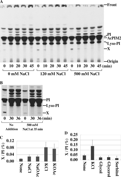
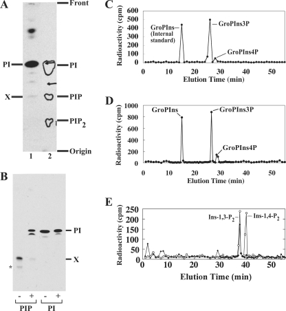
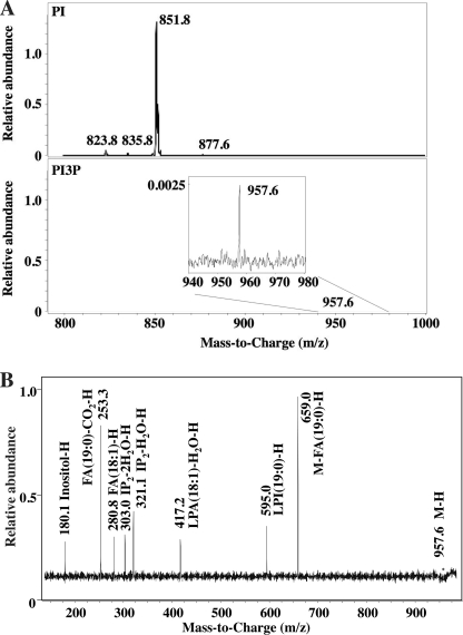
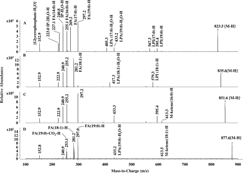
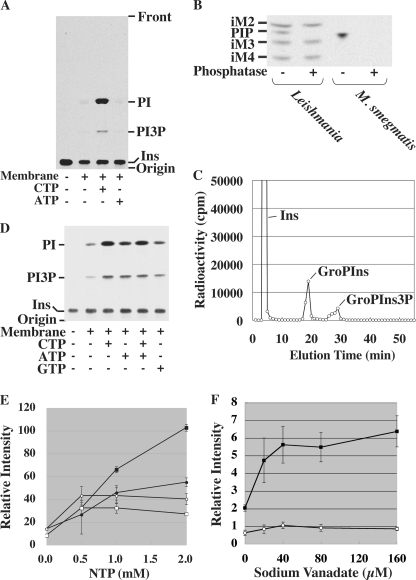
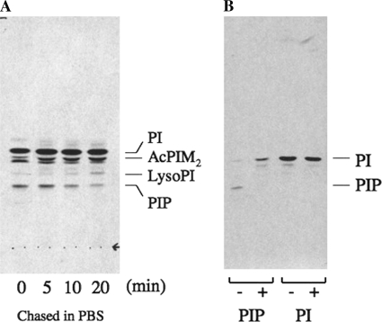
Phosphoinositides play key roles in regulating membrane dynamics and intracellular signaling in eukaryotic cells. However, comparable lipid-based signaling pathways have not been identified in bacteria. Here we show that Mycobacterium smegmatis and other Actinomycetes bacteria can synthesize the phosphoinositide, phosphatidylinositol 3-phosphate (PI3P). This lipid was transiently labeled with [(3)H]inositol. Sensitivity of the purified lipid to alkaline phosphatase, headgroup analysis by high-pressure liquid chromatography, and mass spectrometry demonstrated that it had the structure 1,2-[tuberculostearoyl, octadecenoyl]-sn-glycero 3-phosphoinositol 3-phosphate. Synthesis of PI3P was elevated by salt stress but not by exposure to high concentrations of non-ionic solutes. Synthesis of PI3P in a cell-free system was stimulated by the synthesis of CDP-diacylglycerol, a lipid substrate for phosphatidylinositol (PI) biosynthesis, suggesting that efficient cell-free PI3P synthesis is dependent on de novo PI synthesis. In vitro experiments further indicated that the rapid turnover of this lipid was mediated, at least in part, by a vanadate-sensitive phosphatase. This is the first example of de novo synthesis of PI3P in bacteria, and the transient synthesis in response to environmental stimuli suggests that some bacteria may have evolved similar lipid-mediated signaling pathways to those observed in eukaryotic cells.


Figures

FIGURE 1.
FIGURE 1.
A new [3H]inositol-labeled lipid in M. smegmatis. A, cells were pulse-labeled with [3H]inositol and chased in 25 mm Tris buffer (pH 7.5)/1 mm inositol containing different concentrations of NaCl. AcPIM2, acyl-phosphatidylinositol dimannoside. The radiolabeled species directly above PI is likely to be Ac2PIM1 (35). Other less polar species have not been characterized. B, pulse-chase labeling was conducted in the absence of NaCl as described in A. Cells were then exposed to 500 mm NaCl at 35 min. C and D, cells were labeled with [3H]inositol as in B, and then exposed to increases in the concentration of different salts (300 mm) or non-ionic solutes (600 mm) at 35 min. Cells were harvested after 5 min and label in Lipid X normalized to that in PI. Averages of triplicates with standard deviations are shown.
FIGURE 2.
FIGURE 2.
Structural characterization of Lipid X. A, migration of [3H]inositol-labeled Lipid X (lane 1) on an oxalic acid-impregnated HPTLC plate in comparison to bovine brain phosphoinositides (lane 2). Contours in lane 2 indicate PI, PIP, and PI bisphosphate (PIP2) from brain phosphoinositides, which were visualized by iodine vapor. B, PI and Lipid X were metabolically labeled with [3H]inositol, purified by preparative HPTLC, treated with alkaline phosphatase, and resolved by HPTLC. −, mock; +, alkaline phosphatase. *, a minor contaminant, which may be a PI3P variant. C, [3H]inositol-labeled Lipid X was deacylated, and released GroPIns phosphates were fractionated by HPLC. As an internal standard, GroPIns (generated by deacylating [3H]PI) was coinjected. D, co-elution of mycobacterial Lipid X with the yeast PI3P standard. [3H]Inositol-labeled Lipid X was deacylated as in C. [3H]Inositol-labeled yeast PI/PIP standards were prepared as described under “Experimental Procedures” and deacylated to produce a mixture of GroPIns, GroPIns3P, and GroPIns4P. The yeast standard mixture containing GroPIns (926 cpm), GroPIns3P (355 cpm), and GroPIns4P (206 cpm) was co-injected into the HPLC column with mycobacterial Lipid X (GroPIns3P/GroPIns4P candidates; 570 cpm/66 cpm). Recovery from the yeast GroPIns peak was 90% (830 cpm). E, inositol polyphosphates were generated by deglyceration of the GroPIns3P peak derived from Lipid X and fractionated by HPLC. Open circles, standards; closed diamonds, deglycerated M. smegmatis PI3P headgroup.
FIGURE 3.
FIGURE 3.
Mass spectrometric analysis of PI3P. A, precursor ion scanning MS for molecular species of PI (upper panel) and PI3P (lower panel). B, MS/MS of m/z = 957.6 from PI3P fraction. The mass spectra obtained with different fragmentator voltages and collision energies were merged to form the composite mass spectrum. FA, fatty acid; IP, inositol phosphate; and LPA, lysophosphatidic acid.
FIGURE 4.
FIGURE 4.
MS/MS of M. smegmatis PI species. A–D, fragment ions from m/z = 823.5, m/z = 835.6, m/z = 851.6, m/z = 877.6, respectively. Note that PI from m/z = 823.5 is a mixture of C19:0/C14:0-PI and C17:0/C16:0-PI. The fragmentator was set at 100 V with a collision energy of 45.0 eV and 72 scans. FA, fatty acid; IP, inositol phosphate; LPA, lysophosphatidic acid; and LPI, lyso-PI.
FIGURE 5.
FIGURE 5.
Endogenous PI3P concentration estimated from the relative abundance of synthetic PI3P used as an internal standard. A, the parent (Q1) → product ion (Q3) transition of m/z 848 → 321 was confirmed for the synthetic PI3P standard (C37H73O16P2N1). The fragmentator was set at 100 V with a collision energy of 35.0 or 42.0 eV. The mass spectra obtained with different collision energies were merged to form the composite mass spectrum. B, the PI3P fraction from M. smegmatis (open squares) was spiked with the synthetic PI3P standard (closed circles) at the indicated concentration. Electrospray ionization-MS/MS was used to measure transition pairs in selective multiple reaction monitoring mode. The fragmentator was set at 100 V with a collision energy of 25.0 eV and scanned 200–300 times each.
FIGURE 6.
FIGURE 6.
Synthesis of PI3P in a mycobacterial cell-free system. A, the membrane fraction from a wild-type strain was incubated with [3H]inositol and various nucleotides (2 mm). Ins, inositol. B, PI3P was synthesized in a cell-free system, and a crude lipid extract was treated with 11 units of alkaline phosphatase from bovine intestinal mucosa (Sigma-Aldrich) in a buffer containing 1 m CHES (pH 9.8), 1 mm MgCl2, and 0.008% Triton X-100 at 37 °C for 2 h. Lipid extracts from [3H]inositol-labeled L. mexicana was used as a control, showing that alkaline phosphatase digested PIP specifically without digesting glycoinositol phospholipids (iM2, iM3, and iM4) (22). C, PI3P was prepared by incubating wild-type membranes with [3H]inositol and CTP. Crude lipid extracts were deacylated and applied to an HPLC. Detection of GroPIns3P peak indicated the cell-free synthesis of PI3P in addition to the synthesis of PI (GroPIns peak). In some experiments, as shown here, the elution time of GroPIns was slightly delayed, but GroPIns3P consistently eluted 10–11 min after the elution of GroPIns. Ins, free unincorporated inositol carried over from the cell-free radiolabeling. D, the membrane fraction from the Δino1 mutant was incubated with [3H]inositol and various nucleotides (2 mm). E, reactions were performed as in D with various concentrations of nucleoside triphosphates (NTPs), and plotted as relative radioactivities. Filled symbols, CTP; open symbols, ATP; squares, PI; circles, PI3P. Averages of triplicates with standard deviations are shown. F, the effect of sodium orthovanadate was examined using the system as in D. No nucleotide was added. Averages of triplicates with standard deviations are shown. Open circle, PI; closed square, PI3P.
FIGURE 7.
FIGURE 7.
Synthesis of PIP in C. glutamicum. A, transient synthesis of PIP after chase in phosphate-buffered saline. C. glutamicum was grown to a mid log phase and harvested. The cell pellet was rinsed and incubated in Sauton's minimal medium for 15 min. Radiolabeling with [3H]inositol was performed at 30 °C for 5 min and chased in phosphate-buffered saline for the time indicated. B, alkaline phosphatase treatment of C. glutamicum PIP. PIP or PI was purified from HPTLC plates and subjected to digestion with (+) or without (−) alkaline phosphatase. Note that PIP was converted to a less polar species consistent with PI.

Similar articles

Cited by

References

    1. Whitman M., Downes C. P., Keeler M., Keller T., Cantley L. (1988) Nature 332, 644–646 - PubMed
    1. Kutateladze T. G. (2006) Biochim. Biophys. Acta 1761, 868–877 - PMC - PubMed
    1. Falasca M., Maffucci T. (2006) Arch. Physiol. Biochem. 112, 274–284 - PubMed
    1. Lindmo K., Stenmark H. (2006) J. Cell Sci. 119, 605–614 - PubMed
    1. Engelman J. A., Luo J., Cantley L. C. (2006) Nat. Rev. Genet. 7, 606–619 - PubMed

Publication types