. 2010 May;120(5):1494-505.
doi: 10.1172/JCI38331. Epub 2010 Apr 1.

Myc controls transcriptional regulation of cardiac metabolism and mitochondrial biogenesis in response to pathological stress in mice

Affiliations

Myc controls transcriptional regulation of cardiac metabolism and mitochondrial biogenesis in response to pathological stress in mice

Preeti Ahuja et al. J Clin Invest. 2010 May.

Abstract

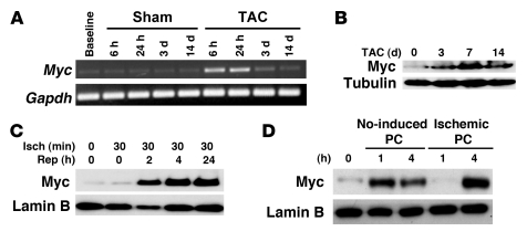
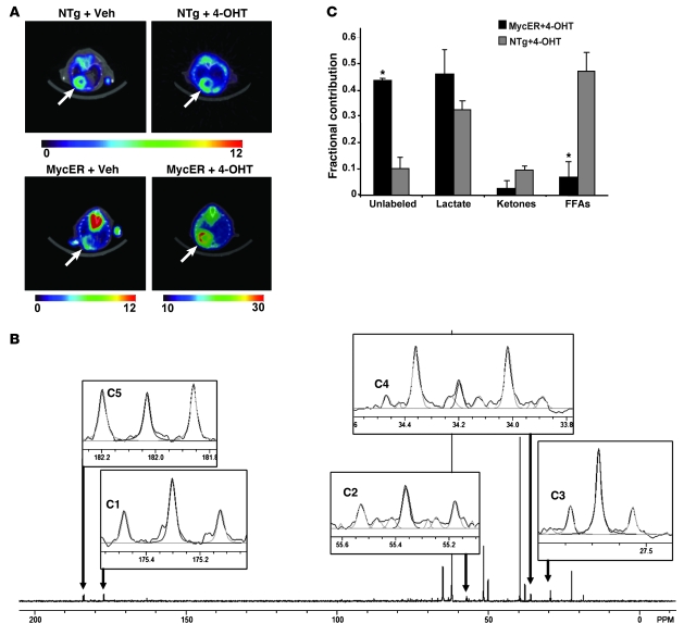
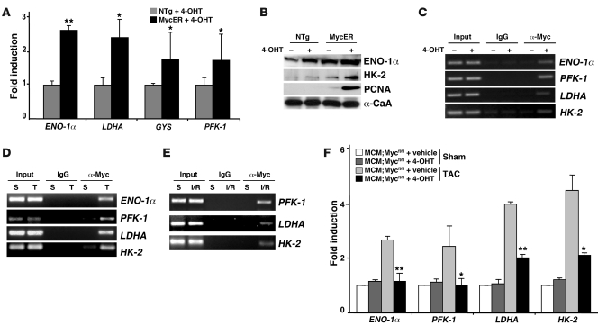
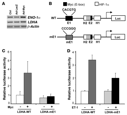
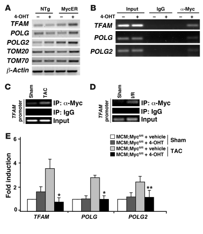
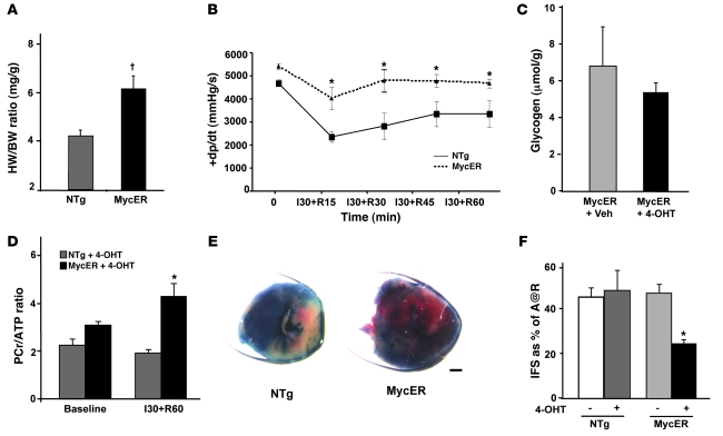
In the adult heart, regulation of fatty acid oxidation and mitochondrial genes is controlled by the PPARgamma coactivator-1 (PGC-1) family of transcriptional coactivators. However, in response to pathological stressors such as hemodynamic load or ischemia, cardiac myocytes downregulate PGC-1 activity and fatty acid oxidation genes in preference for glucose metabolism pathways. Interestingly, despite the reduced PGC-1 activity, these pathological stressors are associated with mitochondrial biogenesis, at least initially. The transcription factors that regulate these changes in the setting of reduced PGC-1 are unknown, but Myc can regulate glucose metabolism and mitochondrial biogenesis during cell proliferation and tumorigenesis in cancer cells. Here we have demonstrated that Myc activation in the myocardium of adult mice increases glucose uptake and utilization, downregulates fatty acid oxidation by reducing PGC-1alpha levels, and induces mitochondrial biogenesis. Inactivation of Myc in the adult myocardium attenuated hypertrophic growth and decreased the expression of glycolytic and mitochondrial biogenesis genes in response to hemodynamic load. Surprisingly, the Myc-orchestrated metabolic alterations were associated with preserved cardiac function and improved recovery from ischemia. Our data suggest that Myc directly regulates glucose metabolism and mitochondrial biogenesis in cardiac myocytes and is an important regulator of energy metabolism in the heart in response to pathologic stress.


Figures

Figure 1
Figure 1. Expression of Myc is upregulated in the heart in response to multiple pathological stressors.
(A) Representative time course of Myc expression in sham or transverse aortic–banded wild-type mice. Semiquantitative PCR was performed on total ventricular RNA. (B) Western blot for expression of Myc in wild-type ventricles after 0 hours, 3 days, 7 days, and 14 days of TAC together with tubulin as a loading control. (C) Temporal expression pattern of Myc protein levels in wild-type hearts as determined by immunoblot analysis at baseline, after 30 minutes of ischemia (Isch), and after 24 hours of reperfusion (Rep). Lamin B was included as loading control. (D) Immunoblotting on ventricular lysates from NO-induced and ischemic preconditioned hearts.
Figure 2
Figure 2. Increased glucose uptake and utilization rates in Myc-activated myocardium.
(A) MicroPET imaging on representative NTg and MycER mice with or without (Veh) 4-OHT treatment (n = 4/group). The relative amount of tracer uptake into the mouse heart 1 hour after intraperitoneal injection of 18F-FDG is indicated by the color scale (range, 0–30). The arrows indicate the cardiac field. (B) Representative NMR spectra from Myc-induced mouse heart. Labeled glutamate carbons are highlighted. Light gray lines represent peak integration with NUTS program. (C) Fractional contribution of acetyl-CoA from the substrates to the citric acid cycle after 4-OHT treatment. *P < 0.05, MycER mice versus NTg littermates; n = 3/group.
Figure 3
Figure 3. Myc directly regulates expression of glycolytic genes in adult myocardium.
(A) Total ventricular RNA from NTg and MycER mice with or without 4-OHT treatment for 7 days assayed by real-time PCR for glycolytic enzymes genes ENO-1α, LDHA, GYS, and PFK-1 in activated MycER versus 4-OHT–treated NTg ventricles (n = 3/group). (B) Total ventricular protein lysates from mice of the indicated genotypes were probed with antibodies against glycolysis-associated proteins (ENO-1α and HK-2) along with α-CaA as a loading control. (C) Myc binds to the glycolytic gene promoter sequences in situ. ChIP analysis performed with anti-Myc antibody and PCR primers to the selected glycolytic genes promoters. Ventricular tissues obtained from MycER mice 24 hours after 4-OHT or vehicle treatment were analyzed. Input lanes show PCR product derived from chromatin before immunoprecipitation to verify equal loading. (D and E) Endogenous Myc binds to the promoters of ENO-1α, PFK-1, LDHA, and HK-2 in response to pathologic stress. Ventricular tissue obtained from wild-type mice subjected to sham (S) operation or TAC (T; 1 day) and after I/R injury (30 minutes of ischemia and 24 hours of reperfusion) were analyzed with anti-Myc antibody and PCR primers to the glycolytic promoters of ENO-1α, PFK-1, LDHA, and HK-2. Input lanes show PCR product derived from chromatin before immunoprecipitation to verify equal loading. (F) Myc-null mice demonstrate an attenuated stress-induced increase in glycolytic genes. Total ventricular RNA from MCM;Mycfl/fl mice with or without 4-OHT treatment subjected to sham or TAC operation was assayed using quantitative real-time PCR for glycolytic genes ENO-1α, PFK-1, LDHA, and HK-2 (n = 3/group). *P < 0.05, **P < 0.001.
Figure 4
Figure 4. Myc is required for induction of the LDHA gene in an E-box–dependent manner in response to hypertrophic agonists.
(A) Total ventricular RNA from NRVMs infected with no virus (Ctrl), AdLacZ, or AdMyc was analyzed by semiquantitative PCR for glycolytic genes (ENO-1α and LDHA) or β-actin control. (B) Schematic diagram depicting the wild-type LDHA promoter containing 2 E-boxes (E1 and E2) and 2 HIF-1α–binding (H1 and H2) sites. The E2 and H2 binding sites overlap. The mutation of the 5ι-E-box, mE1, is also shown. (C) NRVMs were transfected with either LDHA-WT or LDHA-mE1 infected with control or Myc-expressing vector. The relative luciferase activities are shown for the wild-type or mutated LDHA promoter. *P < 0.01 for LDHA-WT + Myc versus LDHA-WT without Myc or LDHA-mE1 + Myc. (D) NRVMs were transfected with either LDHA-WT or mutated promoter LDHA-mE1 and stimulated with vehicle or ET-1 for 16 hours. *P < 0.001 versus LDHA-WT without ET-1; **P < 0.01 versus LDHA-mE1 + ET-1. All data shown are representative of at least 3 independent experiments.
Figure 5
Figure 5. Expression of FAO genes is downregulated with Myc activation in the heart.
(A) Total ventricular RNA from NTg and MycER mice with or without 4-OHT treatment for 7 days assayed by semiquantitative PCR for FAO enzymes genes (PGC-1α, PGC-1β, PPRC, PPARα, ERRα, and NRF-1) together with β-actin as a loading control. (B) Total ventricular protein lysates from mice of the indicated genotypes were probed with antibodies against different FAO associated proteins (PGC-1α and MCAD) along with α-CaA as a loading control. (C) Total ventricular RNA from NTg and MycER mice with or without 4-OHT treatment for 7 days as assayed by semiquantitative PCR for target genes of PPARα and ERRα (CPT-1, MCAD, ATP5A, ATP5B, ATP5C, COX-I, COX-IV, and Cyt-C) together with β-actin as a loading control. (D) Total ventricular protein lysates from mice of the indicated genotypes were probed with antibodies against ATP5A, ATP5B, ATP5C, COX-IV, and α-CaA.
Figure 6
Figure 6. Myc stimulates mitochondrial biogenesis.
(A) Transmission electron microscopy performed on heart sections from NTg and MycER mice with OHT treatment. Scale bar: 1 μm. (B) Total mtDNA isolated from ventricles of NTg and MycER mice with or without 4-OHT treatment and loaded on ethidium bromide–stained agarose gel (1.2%). (C) Quantitative real-time PCR on the mitochondrial gene cytochrome c oxidase subunit I (COX1), along with the nuclear gene PPRC as an internal control from MycER mice with or without 4-OHT treatment. *P < 0.01 versus untreated MycER littermates; n = 3. (D) Representative polarograph of mitochondria isolated from treated NTg and MycER ventricles (n = 4/group) energized with complex I substrates in the presence of 2.5 mM Pi and ADP pulses, at the indicated concentrations. Mitochondria (mit) in both groups responded to ADP additions with transient dissipation of membrane potential (Dy) and acceleration of O2 consumption that slowed down after ADP was phosphorylated and Dy recovered. All data shown are representative of at least 3 independent experiments.
Figure 7
Figure 7. Myc directly regulates genes involved in mitochondrial replication and biogenesis.
(A) RT-PCR analysis performed for TFAM, POLG, POLG2, TOM20, and TOM70 on total RNA prepared from ventricular tissue from NTg and MycER mice with or without 4-OHT treatment. β-Actin was used as a loading control. (B) Myc binds to the TFAM, POLG, and POLG2 promoters in situ. ChIP analysis was performed with anti-Myc antibody and PCR primers to the TFAM, POLG, and POLG2 promoters. Ventricular tissue obtained from MycER mice 24 hours after vehicle or 4-OHT treatment was analyzed. Input lanes show PCR product derived from chromatin before immunoprecipitation to verify equal loading. (C and D) Endogenous Myc binds to the TFAM promoter in response to pathologic stress. Ventricular tissue obtained from wild-type mice subjected to sham operation or TAC (1 day) and after I/R injury (30 minutes of ischemia and 24 hours of reperfusion) were analyzed with anti-Myc antibody and PCR primers to the TFAM promoter. Input lanes show PCR product derived from chromatin before immunoprecipitation to verify equal loading. (E) Myc-null mice demonstrate attenuated stress-induced increase in mitochondrial replication and biogenesis genes. Total ventricular RNA from MCM;Mycfl/fl mice with or without 4-OHT treatment subjected to sham or TAC operation was assayed using quantitative real-time PCR for TFAM, POLG, and POLG2 (n = 3/group). *P < 0.05, **P < 0.001 for vehicle- versus 4-OHT–treated MCM;Mycfl/fl mice after TAC operation.
Figure 8
Figure 8. Myc-activated hearts display improved tolerance to I/R injury.
(A) Activation of MycER increases cardiac mass. Measured heart weights (HW; mg) were normalized to body weight (BW; g); n = 3/group. P < 0.005. (B) Contractile performance of hearts from MycER compared with NTg hearts after OHT treatment measured in Langendorff preparations. Hearts were subjected to 30 minutes of global normothermic ischemia (I) followed by the indicated periods of reperfusion (R). The rate of systolic pressure development (+dp/dt) measured at baseline and over time after reperfusion is shown. *P < 0.05 versus treated NTg littermates; n = 4/group. (C) Glycogen levels in μmol/g of tissue as determined by mass spectrometry analysis on ventricular tissue from MycER mice with or without 4-OHT treatment (n = 3/group). (D) PCr/ATP ratio at baseline and after ischemia for 30 minutes followed by 60 minutes of reperfusion as determined by 31P-NMR on ventricular tissue from NTg and MycER mice after 4-OHT treatment. *P < 0.05 versus treated NTg littermates; n = 4/group. (E) Representative 2,3,5-triphenyltetrazolium chloride–stained hearts from NTg and MycER mice treated with 4-OHT after 24 hours of reperfusion. (F) Mean infarct sizes from NTg and MycER mice with or without 4-OHT treatment after 24 hours of reperfusion are shown. A@R, area at risk. *P < 0.001 versus treated NTg littermates; n = 5/group.

Similar articles

Cited by

References

    1. Lopaschuk GD, Spafford MA. Energy substrate utilization by isolated working hearts from newborn rabbits. Am J Physiol. 1990;258(5 Pt 2):H1274–H1280. - PubMed
    1. Lopaschuk GD, Collins-Nakai RL, Itoi T. Developmental changes in energy substrate use by the heart. Cardiovasc Res. 1992;26(12):1172–1180. doi: 10.1093/cvr/26.12.1172. - DOI - PubMed
    1. Spitkovsky D, et al. Activity of complex III of the mitochondrial electron transport chain is essential for early heart muscle cell differentiation. FASEB J. 2004;18(11):1300–1302. - PubMed
    1. Nadal-Ginard B, Mahdavi V. Molecular basis of cardiac performance. Plasticity of the myocardium generated through protein isoform switches. J Clin Invest. 1989;84(6):1693–1700. doi: 10.1172/JCI114351. - DOI - PMC - PubMed
    1. Schwartz K, Boheler KR, de la Bastie D, Lompre AM, Mercadier JJ. Switches in cardiac muscle gene expression as a result of pressure and volume overload. Am J Physiol. 1992;262(3 pt 2):R364–R369. - PubMed

Publication types