

Review
doi: 10.1172/JCI41303.
Epub 2010 Apr 1.
The key role of vitamin A in spermatogenesis
Affiliations
- PMID: 20364093
- PMCID: PMC2846058
- DOI: 10.1172/JCI41303
Item in Clipboard
Review
The key role of vitamin A in spermatogenesis
J Clin Invest.
2010 Apr.
Abstract
Spermatogenesis in adult mammals is highly organized, with the goal being continual sperm production. Vertebrate testes are arranged into recurring cellular associations that vary with time and distance along the tubule. These changes over time and distance are designated the cycle of the seminiferous epithelium and the spermatogenic wave, respectively. In this Review, we briefly outline the roles that follicle-stimulating hormone (FSH) and testosterone play in regulating spermatogenesis and describe our current understanding of how vitamin A regulates germ cell differentiation and how it may lead to the generation of both the cycle of the seminiferous epithelium and the spermatogenic wave.
Figures

(A) The mammalian testis is composed of seminiferous tubules intertwined so that the “start” and “end” of these tubules are both connected to the rete testis. Immotile sperm flow from the lumen of the seminiferous tubules into the epididymis via the rete testis. During their passage through the epididymis to the vas deferens, sperm acquire their motility. Adapted with permission from Nature Reviews Genetics (59). (B) Histological cross section through an adult mouse testis depicting seminiferous tubules, the peritubular myoid cells, and the interstitium (space between tubules). (C) Expansion of both the undifferentiated (A spermatogonia) and differentiated (A1 spermatogonia) spermatogonial populations occurs by mitosis of these cell types, regulated in part by FSH. Undifferentiated spermatogonia enter the differentiation pathway at the time of the A-to-A1 spermatogonia transition. The red arrow denotes the required action of vitamin A (in the form of RA) in this transition. The subsequent conversion of differentiated spermatogonia to spermatocytes, representing the initiation of meiosis, also requires RA activity. The differentiation of secondary spermatocytes (m2°m) and the process of spermiogenesis (round spermatids to elongated spermatids, then spermatozoa) requires testosterone.
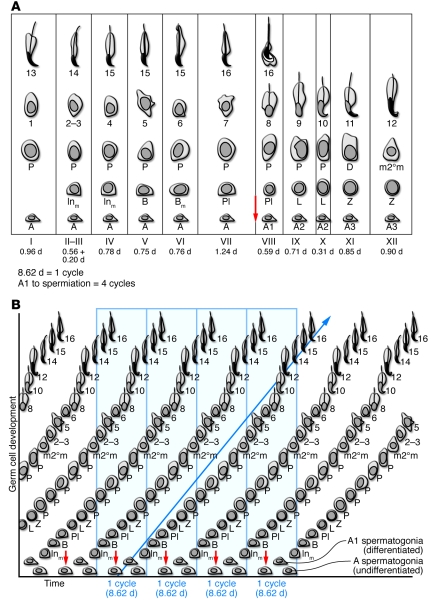
(A) Standard depiction of the cycle of the seminiferous epithelium for the mouse testis. The variable distances between the stages of the cycle are proportional to the duration of each of these cellular associations. The red arrow indicates the time in the cycle when vitamin A is required for the commitment to meiosis. (B) Depiction of how the cycle is generated. Spermatogonia undergo mitotic expansion, and as a result of the action of vitamin A (in the form of RA) (red arrows), they initiate meiosis and ultimately become spermatozoa. The time required for this process from the time of the onset of meiosis to the formation of spermatozoa is particular to the species and the germ cells themselves (blue arrow). The periodic initiation of the differentiation process by vitamin A generates the cellular associations that define the cycle in A. Inm, intermediate (mitosis); B, B spermatogonia; Pl, preleptotene spermatocytes; L, leptotene spermatocytes; Z, zygotene spermatocytes; P, pachytene spermatocytes; D, diplotene spermatocytes; m2°m, secondary spermatocytes. Round and elongating spermatids are labeled as steps 2–3, 8, 12, 16.
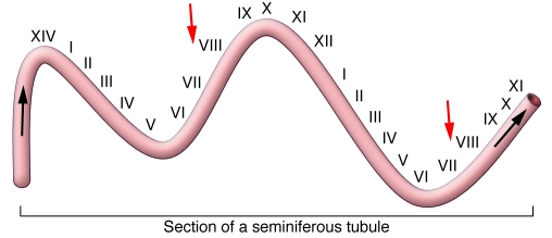
A single seminiferous tubule is depicted, and the stages of the cycle (cellular associations) along the tubule are shown. The spermatogenic wave describes the process in space, while the cycle of the seminiferous epithelium refers to the process in time. The point of meiotic initiation (red arrows) moves along the tubule in the direction of the black arrows. The net result of the wave is the asynchronous (and therefore continual) release of spermatozoa.
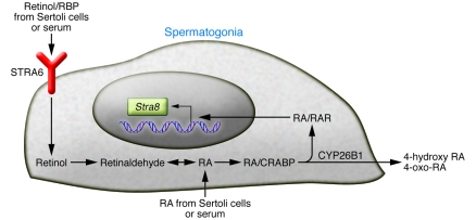
Retinol is delivered to germinal cells on the retinol-binding protein (RBP) and is internalized via the membrane receptor STRA6. It is also plausible that RA is delivered directly to spermatogonia either by Sertoli cells or from the serum. Inside the cell, retinol is converted to RA in a two-step process and can interact with RARs such as RARγ. The RA is bound up by an excess of cellular retinoic acid–binding protein (CRABP). The activated receptor can stimulate transcription of a number of genes including Stra8, which has been shown to be necessary for progression through meiosis. Excess RA can be metabolized by the enzyme CYP26B1 into 4-oxo and 4-hydroxy forms. These forms are then secreted from the cell.
References
-
- de Rooij DG, Russell LD. All you wanted to know about spermatogonia but were afraid to ask. . J Androl. 2000;21(6):776–798. - PubMed
-
- Russell LD, Ettlin, RA, Sinha Hikim AD, Clegg EP.Histological and histopathological evaluation of the testis. Clearwater, FL: Cache River Press; 1990.
-
- Kerr JB, Loveland KL, O’Bryan MK, deKretser DM. Cytology of the testis and intrinsic control mechanisms. In: Neill JD, ed.Physiology of reproduction. New York, NY: Elsevier; 2006:827–947.
-
- Yan HH, Mruk DD, Cheng CY. Junction restructuring and spermatogenesis: the biology, regulation, and implication in male contraceptive development. Curr Top Dev Biol. 2008;80:57–92. - PubMed
Publication types
MeSH terms
Substances
Grants and funding
LinkOut - more resources
Full Text Sources
Other Literature Sources
Medical
